 Movie
Movie Controller

[English] 日本語
 Yorodumi
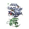
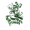
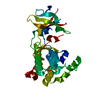
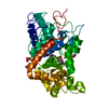
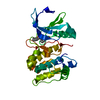
- PDB-1c1y: CRYSTAL STRUCTURE OF RAP.GMPPNP IN COMPLEX WITH THE RAS-BINDING-D... -
+ Open data

- Basic information
| Entry | Database: PDB / ID: 1c1y | ||||||
|---|---|---|---|---|---|---|---|
| Title | CRYSTAL STRUCTURE OF RAP.GMPPNP IN COMPLEX WITH THE RAS-BINDING-DOMAIN OF C-RAF1 KINASE (RAFRBD). | ||||||
 Components Components |
| ||||||
 Keywords Keywords | SIGNALING PROTEIN / GTP-BINDING PROTEINS / PROTEIN-PROTEIN COMPLEX / EFFECTORS | ||||||
| Function / homology |  Function and homology information Function and homology informationpositive regulation of Fc receptor mediated stimulatory signaling pathway / Rap protein signal transduction / regulation of cell junction assembly / death-inducing signaling complex assembly / response to antineoplastic agent / nerve growth factor signaling pathway / intermediate filament cytoskeleton organization / negative regulation of collagen biosynthetic process / positive regulation of vasculogenesis / guanyl-nucleotide exchange factor complex ...positive regulation of Fc receptor mediated stimulatory signaling pathway / Rap protein signal transduction / regulation of cell junction assembly / death-inducing signaling complex assembly / response to antineoplastic agent / nerve growth factor signaling pathway / intermediate filament cytoskeleton organization / negative regulation of collagen biosynthetic process / positive regulation of vasculogenesis / guanyl-nucleotide exchange factor complex / negative regulation of synaptic vesicle exocytosis / ARMS-mediated activation / regulation of Rho protein signal transduction / SHOC2 M1731 mutant abolishes MRAS complex function / Gain-of-function MRAS complexes activate RAF signaling / Rap1 signalling / type B pancreatic cell proliferation / anchoring junction / response to carbohydrate / MET activates RAP1 and RAC1 / insulin secretion involved in cellular response to glucose stimulus / establishment of endothelial barrier / positive regulation of protein kinase activity / Negative feedback regulation of MAPK pathway / IFNG signaling activates MAPKs / Frs2-mediated activation / GP1b-IX-V activation signalling / p130Cas linkage to MAPK signaling for integrins / ERBB2-ERBB3 signaling pathway / neurotrophin TRK receptor signaling pathway / face development / GRB2:SOS provides linkage to MAPK signaling for Integrins / regulation of neurotransmitter receptor localization to postsynaptic specialization membrane / thyroid gland development / pseudopodium / regulation of cell differentiation / somatic stem cell population maintenance / positive regulation of GTPase activity / extrinsic apoptotic signaling pathway via death domain receptors / positive regulation of peptidyl-serine phosphorylation / synaptic vesicle exocytosis / MAP kinase kinase kinase activity / type II interferon-mediated signaling pathway / Schwann cell development / negative regulation of extrinsic apoptotic signaling pathway via death domain receptors / specific granule membrane / negative regulation of protein-containing complex assembly / phagocytic vesicle / response to muscle stretch / Integrin signaling / myelination / CD209 (DC-SIGN) signaling / insulin-like growth factor receptor signaling pathway / liver regeneration / guanyl-nucleotide exchange factor activity / positive regulation of phagocytosis / cellular response to cAMP / thymus development / cellular response to forskolin / adenylate cyclase activator activity / positive regulation of D-glucose import across plasma membrane / small monomeric GTPase / protein localization to plasma membrane / wound healing / RAF activation / cellular response to nerve growth factor stimulus / Signaling by high-kinase activity BRAF mutants / positive regulation of neuron projection development / MAP2K and MAPK activation / small GTPase binding / Stimuli-sensing channels / Signaling by RAF1 mutants / Signaling by moderate kinase activity BRAF mutants / Paradoxical activation of RAF signaling by kinase inactive BRAF / Signaling downstream of RAS mutants / Negative regulation of MAPK pathway / Signaling by BRAF and RAF1 fusions / insulin receptor signaling pathway / cell junction / GDP binding / late endosome / Glucagon-like Peptide-1 (GLP1) regulates insulin secretion / MAPK cascade / nervous system development / presynapse / G protein activity / sperm midpiece / regulation of apoptotic process / protein phosphorylation / early endosome / protein kinase activity / mitochondrial outer membrane / positive regulation of ERK1 and ERK2 cascade / positive regulation of MAPK cascade / non-specific serine/threonine protein kinase / endosome membrane / neuron projection / negative regulation of cell population proliferation / protein serine kinase activity / protein serine/threonine kinase activity Similarity search - Function | ||||||
| Biological species |  Homo sapiens (human) Homo sapiens (human) | ||||||
| Method |  X-RAY DIFFRACTION / SYNCHROTRON / Resolution: 1.9 Å X-RAY DIFFRACTION / SYNCHROTRON / Resolution: 1.9 Å | ||||||
 Authors Authors | Nassar, N. | ||||||
 Citation Citation | Journal: Nature / Year: 1995 Title: The 2.2 A crystal structure of the Ras-binding domain of the serine/threonine kinase c-Raf1 in complex with Rap1A and a GTP analogue. Authors: Nassar, N. #1: Journal: Nat.Struct.Biol. / Year: 1996Title: Ras/Rap effector specificty determined by charge reversal. Authors: Nassar, N. #2: Journal: Nature / Year: 2008Title: Structure of Epac2 in complex with a cyclic AMP analogue and RAP1B. Authors: Holger Rehmann / Ernesto Arias-Palomo / Michael A Hadders / Frank Schwede / Oscar Llorca / Johannes L Bos /  Abstract: Epac proteins are activated by binding of the second messenger cAMP and then act as guanine nucleotide exchange factors for Rap proteins. The Epac proteins are involved in the regulation of cell ...Epac proteins are activated by binding of the second messenger cAMP and then act as guanine nucleotide exchange factors for Rap proteins. The Epac proteins are involved in the regulation of cell adhesion and insulin secretion. Here we have determined the structure of Epac2 in complex with a cAMP analogue (Sp-cAMPS) and RAP1B by X-ray crystallography and single particle electron microscopy. The structure represents the cAMP activated state of the Epac2 protein with the RAP1B protein trapped in the course of the exchange reaction. Comparison with the inactive conformation reveals that cAMP binding causes conformational changes that allow the cyclic nucleotide binding domain to swing from a position blocking the Rap binding site towards a docking site at the Ras exchange motif domain. | ||||||
| History |
|
- Structure visualization
| Structure viewer | Molecule: MolmilJmol/JSmol |
|---|
- Downloads & links
-Download
| PDBx/mmCIF format | 1c1y.cif.gz | 67.9 KB | Display | PDBx/mmCIF format |
|---|---|---|---|---|
| PDB format | pdb1c1y.ent.gz | 49.3 KB | Display | PDB format |
| PDBx/mmJSON format | 1c1y.json.gz | Tree view | PDBx/mmJSON format | |
| Others |  Other downloads Other downloads |
-Validation report
| Arichive directory | https://data.pdbj.org/pub/pdb/validation_reports/c1/1c1yftp://data.pdbj.org/pub/pdb/validation_reports/c1/1c1y | HTTPS FTP |
|---|
-Related structure data
| Related structure data | |
|---|---|
| Similar structure data |


-Links
 PDBj
PDBj


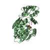
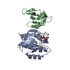
- Assembly
| Deposited unit | 
| ||||||||
|---|---|---|---|---|---|---|---|---|---|
| 1 |
| ||||||||
| Unit cell |
|
-Components
-Protein , 2 types, 2 molecules AB
| #1: Protein | Mass: 19055.641 Da / Num. of mol.: 1 / Fragment: RAP, RESIDUES 1-167 Source method: isolated from a genetically manipulated source Source: (gene. exp.) Homo sapiens (human) / Plasmid: PBKS / Production host:  |
|---|---|
| #2: Protein | Mass: 8789.290 Da / Num. of mol.: 1 / Fragment: RAFRBD, RESIDUES 51-131 Source method: isolated from a genetically manipulated source Source: (gene. exp.) Homo sapiens (human) / Plasmid: PGEX-2T / Production host: References: UniProt: P04049, Transferases; Transferring phosphorus-containing groups; Phosphotransferases with an alcohol group as acceptor |
-Non-polymers , 4 types, 86 molecules 


| #3: Chemical | ChemComp-MG /  Mass: 24.305 Da / Num. of mol.: 1 / Source method: obtained synthetically / Formula: Mg Mass: 24.305 Da / Num. of mol.: 1 / Source method: obtained synthetically / Formula: Mg |
|---|---|
| #4: Chemical | ChemComp-GTP /  Mass: 523.180 Da / Num. of mol.: 1 / Source method: obtained synthetically / Formula: C10H16N5O14P3 / Comment: GTP, energy-carrying molecule*YM Mass: 523.180 Da / Num. of mol.: 1 / Source method: obtained synthetically / Formula: C10H16N5O14P3 / Comment: GTP, energy-carrying molecule*YM |
| #5: Chemical | ChemComp-CA /  Mass: 40.078 Da / Num. of mol.: 1 / Source method: obtained synthetically / Formula: Ca Mass: 40.078 Da / Num. of mol.: 1 / Source method: obtained synthetically / Formula: Ca |
| #6: Water | ChemComp-HOH / Mass: 18.015 Da / Num. of mol.: 83 / Source method: isolated from a natural source / Formula: H2O |
-Experimental details
-Experiment
| Experiment | Method: X-RAY DIFFRACTION / Number of used crystals: 1 |
|---|
- Sample preparation
| Crystal | Density Matthews: 2.86 Å3/Da / Density % sol: 56.93 % | ||||||||||||||||||||||||||||||||||||||||||||||||||||||||||||||||||||||||||||||||||||
|---|---|---|---|---|---|---|---|---|---|---|---|---|---|---|---|---|---|---|---|---|---|---|---|---|---|---|---|---|---|---|---|---|---|---|---|---|---|---|---|---|---|---|---|---|---|---|---|---|---|---|---|---|---|---|---|---|---|---|---|---|---|---|---|---|---|---|---|---|---|---|---|---|---|---|---|---|---|---|---|---|---|---|---|---|---|
| Crystal grow | Temperature: 300 K / Method: vapor diffusion, hanging drop / pH: 7.6 Details: mmePeg5000, magnesium chloride, calcium chloride, ammonium sulphate., pH 7.6, VAPOR DIFFUSION, HANGING DROP, temperature 300K | ||||||||||||||||||||||||||||||||||||||||||||||||||||||||||||||||||||||||||||||||||||
| Crystal grow | *PLUS | ||||||||||||||||||||||||||||||||||||||||||||||||||||||||||||||||||||||||||||||||||||
| Components of the solutions | *PLUS
|
-Data collection
| Diffraction | Mean temperature: 278 K |
|---|---|
| Diffraction source | Source: SYNCHROTRON / Site: EMBL/DESY, HAMBURG  / Beamline: BW7B / Wavelength: 0.87 / Beamline: BW7B / Wavelength: 0.87 |
| Detector | Type: MARRESEARCH / Detector: IMAGE PLATE / Date: Sep 13, 1994 |
| Radiation | Protocol: SINGLE WAVELENGTH / Monochromatic (M) / Laue (L): M / Scattering type: x-ray |
| Radiation wavelength | Wavelength: 0.87 Å / Relative weight: 1 |
| Reflection | Resolution: 1.9→20 Å / Num. all: 24787 / Num. obs: 24787 / % possible obs: 98 % / Observed criterion σ(I): 2 / Redundancy: 4.3 % / Biso Wilson estimate: 24.2 Å2 / Rmerge(I) obs: 0.038 / Net I/σ(I): 12 |
| Reflection shell | Resolution: 1.9→2.02 Å / Redundancy: 3 % / Rmerge(I) obs: 0.325 / Num. unique all: 3569 / % possible all: 93.6 |
| Reflection | *PLUS % possible obs: 98 % |
- Processing
| Software |
| ||||||||||||||||||||||||||||||||||||||||
|---|---|---|---|---|---|---|---|---|---|---|---|---|---|---|---|---|---|---|---|---|---|---|---|---|---|---|---|---|---|---|---|---|---|---|---|---|---|---|---|---|---|
| Refinement | Resolution: 1.9→20 Å / Rfactor Rfree error: 0.005 / Data cutoff high absF: 7734287.57 / Data cutoff low absF: 0 / Isotropic thermal model: RESTRAINED / Cross valid method: THROUGHOUT / σ(F): 0 / Stereochemistry target values: Engh & Huber / Details: Maximum Likelyhood refinement using CNS_0.4.
| ||||||||||||||||||||||||||||||||||||||||
| Solvent computation | Solvent model: FLAT MODEL / Bsol: 37.03 Å2 / ksol: 0.332 e/Å3 | ||||||||||||||||||||||||||||||||||||||||
| Displacement parameters | Biso mean: 38.8 Å2
| ||||||||||||||||||||||||||||||||||||||||
| Refine analyze |
| ||||||||||||||||||||||||||||||||||||||||
| Refinement step | Cycle: LAST / Resolution: 1.9→20 Å
| ||||||||||||||||||||||||||||||||||||||||
| Refine LS restraints |
| ||||||||||||||||||||||||||||||||||||||||
| LS refinement shell | Resolution: 1.9→2.02 Å / Rfactor Rfree error: 0.019 / Total num. of bins used: 6
| ||||||||||||||||||||||||||||||||||||||||
| Xplor file |
| ||||||||||||||||||||||||||||||||||||||||
| Software | *PLUS Name: CNS / Version: 0.4 / Classification: refinement | ||||||||||||||||||||||||||||||||||||||||
| Refinement | *PLUS σ(F): 0 / % reflection Rfree: 9.9 % | ||||||||||||||||||||||||||||||||||||||||
| Solvent computation | *PLUS | ||||||||||||||||||||||||||||||||||||||||
| Displacement parameters | *PLUS Biso mean: 38.8 Å2 | ||||||||||||||||||||||||||||||||||||||||
| Refine LS restraints | *PLUS
| ||||||||||||||||||||||||||||||||||||||||
| LS refinement shell | *PLUS Rfactor Rfree: 0.38 / % reflection Rfree: 9.9 % / Rfactor Rwork: 0.357 |
