negative regulation of cell-cell adhesion mediated by integrin / monoatomic cation homeostasis / chemokine (C-C motif) ligand 21 signaling pathway / lymphocyte migration into lymph node / positive regulation of termination of RNA polymerase II transcription / negative regulation of pancreatic juice secretion / protein insertion into ER membrane by stop-transfer membrane-anchor sequence / negative regulation of small GTPase mediated signal transduction / negative regulation of sodium ion transport / positive regulation of mitotic cytokinesis ...negative regulation of cell-cell adhesion mediated by integrin / monoatomic cation homeostasis / chemokine (C-C motif) ligand 21 signaling pathway / lymphocyte migration into lymph node / positive regulation of termination of RNA polymerase II transcription / negative regulation of pancreatic juice secretion / protein insertion into ER membrane by stop-transfer membrane-anchor sequence / negative regulation of small GTPase mediated signal transduction / negative regulation of sodium ion transport / positive regulation of mitotic cytokinesis / intracellular membraneless organelle / negative regulation of heterotypic cell-cell adhesion / regulation of mRNA export from nucleus / intracellular chloride ion homeostasis / positive regulation of T cell chemotaxis / regulation of sodium ion transmembrane transport / cellular response to chemokine / negative regulation of leukocyte cell-cell adhesion / potassium ion homeostasis / cellular hyperosmotic response / membraneless organelle assembly / cell volume homeostasis / regulation of monoatomic cation transmembrane transport / positive regulation of systemic arterial blood pressure / protein kinase activator activity / negative regulation of protein localization to plasma membrane / neuron development / phosphatase binding / regulation of sodium ion transport / monoatomic ion transport / negative regulation of protein ubiquitination / negative regulation of autophagy / sodium ion transmembrane transport / molecular condensate scaffold activity / Stimuli-sensing channels / positive regulation of angiogenesis / mitotic spindle / positive regulation of canonical Wnt signaling pathway / T cell receptor signaling pathway / heart development / protein phosphorylation / protein kinase activity / non-specific serine/threonine protein kinase / intracellular signal transduction / protein serine kinase activity / protein serine/threonine kinase activity / DNA damage response / protein kinase binding / signal transduction / protein-containing complex / ATP binding / membrane / nucleus / cytoplasm / cytosol Similarity search - Function
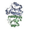
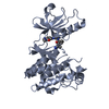
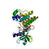
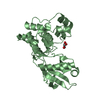
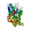
Serine/threonine-proteinkinaseWNK1 / Erythrocyte 65 kDa protein / p65 / Kinase deficient protein / Protein kinase lysine-deficient 1 / ...Erythrocyte 65 kDa protein / p65 / Kinase deficient protein / Protein kinase lysine-deficient 1 / Protein kinase with no lysine 1 / hWNK1
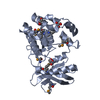
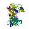
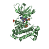
Mass: 31538.545 Da / Num. of mol.: 1 / Fragment: Serine/Threonine Protein kinase WNK1 Source method: isolated from a genetically manipulated source Source: (gene. exp.) Homo sapiens (human) / Gene: WNK1, HSN2, KDP, KIAA0344, PRKWNK1 / Production host: Escherichia coli (E. coli) References: UniProt: Q9H4A3, non-specific serine/threonine protein kinase
In the structure databanks used in Yorodumi, some data are registered as the other names, "COVID-19 virus" and "2019-nCoV". Here are the details of the virus and the list of structure data.
Jan 31, 2019. EMDB accession codes are about to change! (news from PDBe EMDB page)
EMDB accession codes are about to change! (news from PDBe EMDB page)
The allocation of 4 digits for EMDB accession codes will soon come to an end. Whilst these codes will remain in use, new EMDB accession codes will include an additional digit and will expand incrementally as the available range of codes is exhausted. The current 4-digit format prefixed with “EMD-” (i.e. EMD-XXXX) will advance to a 5-digit format (i.e. EMD-XXXXX), and so on. It is currently estimated that the 4-digit codes will be depleted around Spring 2019, at which point the 5-digit format will come into force.
The EM Navigator/Yorodumi systems omit the EMD- prefix.
Related info.:Q: What is EMD? / ID/Accession-code notation in Yorodumi/EM Navigator
Yorodumi is a browser for structure data from EMDB, PDB, SASBDB, etc.
This page is also the successor to EM Navigator detail page, and also detail information page/front-end page for Omokage search.
The word "yorodu" (or yorozu) is an old Japanese word meaning "ten thousand". "mi" (miru) is to see.
Related info.:EMDB / PDB / SASBDB / Comparison of 3 databanks / Yorodumi Search / Aug 31, 2016. New EM Navigator & Yorodumi / Yorodumi Papers / Jmol/JSmol / Function and homology information / Changes in new EM Navigator and Yorodumi
 Movie
Movie Controller


 Open data
 Basic information
 Components
 Keywords
 Function and homology information
 Homo sapiens (human)
 X-RAY DIFFRACTION /
  Authors
 Citation
 Structure visualization
 Downloads & links
 Other downloads
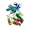


 PDBj
PDBj Assembly
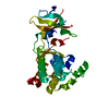


 Mass: 94.971 Da / Num. of mol.: 1 / Source method: obtained synthetically / Formula: PO4
 Mass: 18.015 Da / Num. of mol.: 234 / Source method: isolated from a natural source / Formula: H2O
 Sample preparation
 / Beamline: 19-ID / Wavelength: 1 Å
 Processing