 Movie
Movie Controller


+ Open data

- Basic information
| Entry | Database: PDB / ID: 1c03 | ||||||
|---|---|---|---|---|---|---|---|
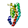
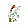
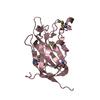
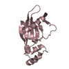
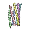
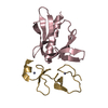
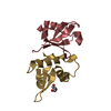
| Title | CRYSTAL STRUCTURE OF YPD1P (TRICLINIC FORM) | ||||||
 Components Components | HYPOTHETICAL PROTEIN YDL235C | ||||||
 Keywords Keywords | SIGNALING PROTEIN / FOUR HELICAL BUNDLE / SIGNAL TRANSDUCTION | ||||||
| Function / homology |  Function and homology information Function and homology informationtransferase activity, transferring phosphorus-containing groups / protein histidine kinase binding / histidine phosphotransfer kinase activity / osmosensory signaling via phosphorelay pathway / phosphorelay signal transduction system / nucleus / cytoplasm Similarity search - Function | ||||||
| Biological species |  | ||||||
| Method |  X-RAY DIFFRACTION / SYNCHROTRON / Resolution: 2.3 Å X-RAY DIFFRACTION / SYNCHROTRON / Resolution: 2.3 Å | ||||||
 Authors Authors | Song, H.K. / Lee, J.Y. / Lee, M.G. / Suh, S.W. | ||||||
 Citation Citation | Journal: J.Mol.Biol. / Year: 1999 Title: Insights into eukaryotic multistep phosphorelay signal transduction revealed by the crystal structure of Ypd1p from Saccharomyces cerevisiae. Authors: Song, H.K. / Lee, J.Y. / Lee, M.G. / Moon, J. / Min, K. / Yang, J.K. / Suh, S.W. #1: Journal: Acta Crystallogr.,Sect.D / Year: 1999Title: Crystallization and preliminary X-ray analysis of Saccharomyces cerevisiae Ypd1p, a key intermediate in phosphorelay signal transduction Authors: Lee, M.G. / Lee, J.Y. / Song, H.K. / Suh, S.W. | ||||||
| History |
|
- Structure visualization
| Structure viewer | Molecule: MolmilJmol/JSmol |
|---|
- Downloads & links
-Download
| PDBx/mmCIF format | 1c03.cif.gz | 137.3 KB | Display | PDBx/mmCIF format |
|---|---|---|---|---|
| PDB format | pdb1c03.ent.gz | 111 KB | Display | PDB format |
| PDBx/mmJSON format | 1c03.json.gz | Tree view | PDBx/mmJSON format | |
| Others |  Other downloads Other downloads |
-Validation report
| Arichive directory | https://data.pdbj.org/pub/pdb/validation_reports/c0/1c03ftp://data.pdbj.org/pub/pdb/validation_reports/c0/1c03 | HTTPS FTP |
|---|
-Related structure data


-Links
 PDBj
PDBj- Assembly
| Deposited unit | 
| ||||||||||
|---|---|---|---|---|---|---|---|---|---|---|---|
| 1 | 
| ||||||||||
| 2 | 
| ||||||||||
| 3 | 
| ||||||||||
| 4 | 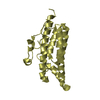
| ||||||||||
| Unit cell |
|
-Components
| #1: Protein | Mass: 19325.799 Da / Num. of mol.: 4 / Mutation: C-TERMINAL 6 HIS-TAG Source method: isolated from a genetically manipulated source Details: TRICLINIC FORM Source: (gene. exp.) Plasmid: PET-22B / Production host:  #2: Water | ChemComp-HOH / |  Mass: 18.015 Da / Num. of mol.: 176 / Source method: isolated from a natural source / Formula: H2O Mass: 18.015 Da / Num. of mol.: 176 / Source method: isolated from a natural source / Formula: H2O |
|---|
-Experimental details
-Experiment
| Experiment | Method: X-RAY DIFFRACTION / Number of used crystals: 1 |
|---|
- Sample preparation
| Crystal | Density Matthews: 2.9 Å3/Da / Density % sol: 57.65 % |
|---|---|
| Crystal grow | Temperature: 297 K / Method: vapor diffusion, hanging drop / pH: 7.36 Details: PEG 400, ammonium sulfate, Hepes, Li2SO4, pH 7.36, VAPOR DIFFUSION, HANGING DROP, temperature 297K |
| Crystal grow | *PLUS Temperature: 296 K |
| Components of the solutions | *PLUS Conc.: 23 mg/ml / Common name: protein |
-Data collection
| Diffraction | Mean temperature: 293 K |
|---|---|
| Diffraction source | Source: SYNCHROTRON / Site: Photon Factory  / Beamline: BL-6A / Wavelength: 1 / Beamline: BL-6A / Wavelength: 1 |
| Detector | Type: FUJI / Detector: IMAGE PLATE / Date: Nov 20, 1998 |
| Radiation | Protocol: SINGLE WAVELENGTH / Monochromatic (M) / Laue (L): M / Scattering type: x-ray |
| Radiation wavelength | Wavelength: 1 Å / Relative weight: 1 |
| Reflection | Resolution: 2.3→50 Å / Num. all: 59932 / Num. obs: 59932 / % possible obs: 88.3 % / Observed criterion σ(F): 0 / Observed criterion σ(I): 0 / Redundancy: 1.7 % / Biso Wilson estimate: 35 Å2 / Rmerge(I) obs: 0.044 / Net I/σ(I): 12.9 |
| Reflection shell | Resolution: 2.3→2.4 Å / Redundancy: 1.6 % / Rmerge(I) obs: 0.26 / % possible all: 85.9 |
| Reflection | *PLUS Num. obs: 35254 |
| Reflection shell | *PLUS % possible obs: 85.9 % |
- Processing
| Software |
| |||||||||||||||||||||||||
|---|---|---|---|---|---|---|---|---|---|---|---|---|---|---|---|---|---|---|---|---|---|---|---|---|---|---|
| Refinement | Resolution: 2.3→8 Å / σ(F): 0 / σ(I): 0 / Stereochemistry target values: Engh & Huber
| |||||||||||||||||||||||||
| Refinement step | Cycle: LAST / Resolution: 2.3→8 Å
| |||||||||||||||||||||||||
| Refine LS restraints |
|
