 Movie
Movie Controller

[English] 日本語
 Yorodumi
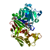
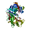
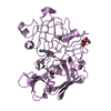
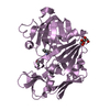
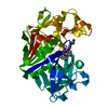
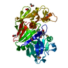
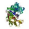
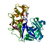
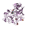
- PDB-1bxq: ACID PROTEINASE (PENICILLOPEPSIN) COMPLEX WITH PHOSPHONATE INHIBITOR. -
+ Open data

- Basic information
| Entry | Database: PDB / ID: 1bxq | ||||||
|---|---|---|---|---|---|---|---|
| Title | ACID PROTEINASE (PENICILLOPEPSIN) COMPLEX WITH PHOSPHONATE INHIBITOR. | ||||||
 Components Components | PROTEIN (PENICILLOPEPSIN) | ||||||
 Keywords Keywords | HYDROLASE / PHOSPHONATE INHIBITORS | ||||||
| Function / homology |  Function and homology information Function and homology informationpenicillopepsin / aspartic-type endopeptidase activity / proteolysis / extracellular region Similarity search - Function | ||||||
| Biological species |  Penicillium janthinellum (fungus) Penicillium janthinellum (fungus) | ||||||
| Method |  X-RAY DIFFRACTION / OTHER / Resolution: 1.41 Å X-RAY DIFFRACTION / OTHER / Resolution: 1.41 Å | ||||||
 Authors Authors | Parrish, J.C. / Khan, A.R. / Fraser, M.E. / Smith, W.W. / Bartlett, P.A. / James, M.N.G. | ||||||
 Citation Citation | Journal: Biochemistry / Year: 1998 Title: Lowering the entropic barrier for binding conformationally flexible inhibitors to enzymes. Authors: Khan, A.R. / Parrish, J.C. / Fraser, M.E. / Smith, W.W. / Bartlett, P.A. / James, M.N. #1: Journal: J.Am.Chem.Soc. / Year: 1998Title: Macrocyclic Inhibitors of Penicillopepsin. 3. Design, Synthesis, and Evaluation of an Inhibitor Bridged between P2 and P1' Authors: Smith, W.W. / Bartlett, P.A. #2: Journal: J.Am.Chem.Soc. / Year: 1998Title: Macrocyclic Inhibitors of Penicillopepsin. 2.X-Ray Crystallographic Analyses of Penicillopepsin Complexed with a P3-P1 Macrocyclic Peptidyl Inhibitor and with its Two Acyclic Analogues Authors: Ding, J. / Fraser, M.E. / Meyer, J.H. / Bartlett, P.A. / James, M.N.G. | ||||||
| History |
|
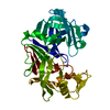
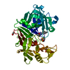
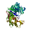
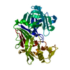
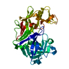
- Structure visualization
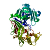
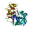
| Structure viewer | Molecule: MolmilJmol/JSmol |
|---|
- Downloads & links
-Download
| PDBx/mmCIF format | 1bxq.cif.gz | 84.4 KB | Display | PDBx/mmCIF format |
|---|---|---|---|---|
| PDB format | pdb1bxq.ent.gz | 62 KB | Display | PDB format |
| PDBx/mmJSON format | 1bxq.json.gz | Tree view | PDBx/mmJSON format | |
| Others |  Other downloads Other downloads |
-Validation report
| Arichive directory | https://data.pdbj.org/pub/pdb/validation_reports/bx/1bxqftp://data.pdbj.org/pub/pdb/validation_reports/bx/1bxq | HTTPS FTP |
|---|
-Related structure data
| Related structure data | 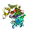 1bxoC 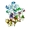 1pplS S: Starting model for refinement C: citing same article ( |
|---|---|
| Similar structure data |


-Links
 PDBj
PDBj


- Assembly
| Deposited unit | 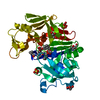
| ||||||||||||
|---|---|---|---|---|---|---|---|---|---|---|---|---|---|
| 1 |
| ||||||||||||
| Unit cell |
| ||||||||||||
| Components on special symmetry positions |
|
-Components
-Protein / Sugars , 2 types, 3 molecules A
| #1: Protein | Mass: 33468.809 Da / Num. of mol.: 1 / Source method: isolated from a natural source / Source: (natural) Penicillium janthinellum (fungus) / References: UniProt: P00798, penicillopepsin |
|---|---|
| #2: Sugar |  Type: D-saccharide, alpha linking / Mass: 180.156 Da / Num. of mol.: 2 Type: D-saccharide, alpha linking / Mass: 180.156 Da / Num. of mol.: 2Source method: isolated from a genetically manipulated source Formula: C6H12O6 |
-Non-polymers , 5 types, 412 molecules 

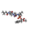


| #3: Chemical |  Mass: 96.063 Da / Num. of mol.: 2 / Source method: obtained synthetically / Formula: SO4 Mass: 96.063 Da / Num. of mol.: 2 / Source method: obtained synthetically / Formula: SO4#4: Chemical | ChemComp-ACT / |  Mass: 59.044 Da / Num. of mol.: 1 / Source method: obtained synthetically / Formula: C2H3O2 Mass: 59.044 Da / Num. of mol.: 1 / Source method: obtained synthetically / Formula: C2H3O2#5: Chemical | ChemComp-PP8 / |  Mass: 626.679 Da / Num. of mol.: 1 / Source method: obtained synthetically / Formula: C29H47N4O9P Mass: 626.679 Da / Num. of mol.: 1 / Source method: obtained synthetically / Formula: C29H47N4O9P#6: Chemical |  Mass: 92.094 Da / Num. of mol.: 3 / Source method: obtained synthetically / Formula: C3H8O3 Mass: 92.094 Da / Num. of mol.: 3 / Source method: obtained synthetically / Formula: C3H8O3#7: Water | ChemComp-HOH / | Mass: 18.015 Da / Num. of mol.: 405 / Source method: isolated from a natural source / Formula: H2O |
|---|
-Details
| Has protein modification | Y |
|---|
-Experimental details
-Experiment
| Experiment | Method: X-RAY DIFFRACTION / Number of used crystals: 1 |
|---|
- Sample preparation
| Crystal | Density Matthews: 1.51 Å3/Da / Density % sol: 18.4 % | ||||||||||||||||||||
|---|---|---|---|---|---|---|---|---|---|---|---|---|---|---|---|---|---|---|---|---|---|
| Crystal grow | pH: 4.6 / Details: 0.1 M CH3COONA 35% SATD. AMMONIUM SULPHATE PH 4.6 | ||||||||||||||||||||
| Crystal grow | *PLUS Method: vapor diffusion, hanging drop | ||||||||||||||||||||
| Components of the solutions | *PLUS
|
-Data collection
| Diffraction | Mean temperature: 100 K |
|---|---|
| Diffraction source | Source: ROTATING ANODE / Wavelength: 1.5418 |
| Detector | Type: SIEMENS / Detector: AREA DETECTOR / Date: Feb 15, 1998 |
| Radiation | Monochromator: GRAPHITE / Protocol: SINGLE WAVELENGTH / Monochromatic (M) / Laue (L): M / Scattering type: x-ray |
| Radiation wavelength | Wavelength: 1.5418 Å / Relative weight: 1 |
| Reflection | Resolution: 1.44→8 Å / Num. obs: 45997 / % possible obs: 94.8 % / Redundancy: 2.33 % / Rmerge(I) obs: 0.074 / Net I/σ(I): 56.035 |
| Reflection shell | Resolution: 1.45→1.49 Å / Redundancy: 1.31 % / Rmerge(I) obs: 0.0938 / Mean I/σ(I) obs: 13.26 / % possible all: 66.3 |
| Reflection | *PLUS Num. measured all: 107304 |
| Reflection shell | *PLUS % possible obs: 66.3 % |
- Processing
| Software |
| |||||||||||||||||||||||||||||||||
|---|---|---|---|---|---|---|---|---|---|---|---|---|---|---|---|---|---|---|---|---|---|---|---|---|---|---|---|---|---|---|---|---|---|---|
| Refinement | Method to determine structure: OTHER Starting model: 1PPL Resolution: 1.41→10 Å / Num. parameters: 11619 / Num. restraintsaints: 10360 / Cross valid method: FREE R / σ(F): 0 / Stereochemistry target values: ENGH AND HUBER
| |||||||||||||||||||||||||||||||||
| Solvent computation | Solvent model: MOEWS & KRETSINGER | |||||||||||||||||||||||||||||||||
| Refine analyze | Num. disordered residues: 10 / Occupancy sum hydrogen: 2279.3 / Occupancy sum non hydrogen: 2861.7 | |||||||||||||||||||||||||||||||||
| Refinement step | Cycle: LAST / Resolution: 1.41→10 Å
| |||||||||||||||||||||||||||||||||
| Refine LS restraints |
|
