 Movie
Movie Controller

[English] 日本語
 Yorodumi
- PDB-1apt: CRYSTALLOGRAPHIC ANALYSIS OF A PEPSTATIN ANALOGUE BINDING TO THE ... -
+ Open data

- Basic information
| Entry | Database: PDB / ID: 1apt | ||||||
|---|---|---|---|---|---|---|---|
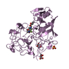
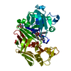
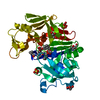
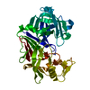
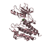
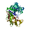
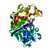
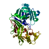
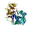
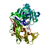
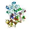
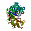
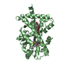
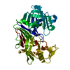
| Title | CRYSTALLOGRAPHIC ANALYSIS OF A PEPSTATIN ANALOGUE BINDING TO THE ASPARTYL PROTEINASE PENICILLOPEPSIN AT 1.8 ANGSTROMS RESOLUTION | ||||||
 Components Components |
| ||||||
 Keywords Keywords | HYDROLASE/HYDROLASE INHIBITOR / ACID PROTEINASE / HYDROLASE-HYDROLASE INHIBITOR complex | ||||||
| Function / homology |  Function and homology information Function and homology informationpenicillopepsin / aspartic-type endopeptidase activity / proteolysis / extracellular region Similarity search - Function | ||||||
| Biological species |  Penicillium janthinellum (fungus) Penicillium janthinellum (fungus) | ||||||
| Method |  X-RAY DIFFRACTION / Resolution: 1.8 Å X-RAY DIFFRACTION / Resolution: 1.8 Å | ||||||
 Authors Authors | Sielecki, A.R. / James, M.N.G. | ||||||
 Citation Citation | Journal: Peptides: Structure and Function, Proceedings of the of the Eighth American Peptide Symposium Year: 1983 Title: Crystallographic Analysis of a Pepstatin Analogue Binding to the Aspartyl Proteinase Penicillopepsin at 1.8 Angstroms Resolution Authors: James, M.N.G. / Sielecki, A.R. / Moult, J. #1: Journal: Biochemistry / Year: 1992Title: Crystallographic Analysis of Transition State Mimics Bound to Penicillopepsin: Difluorostatine-and Difluorostatone-Containing Peptides Authors: James, M.N.G. / Sielecki, A.R. / Hayakawa, K. / Gelb, M.H. #2: Journal: Biological Macromolecules and Assemblies / Year: 1987Title: Aspartic Proteinases and Their Catalytic Pathway Authors: James, M.N.G. / Sielecki, A.R. #3: Journal: Biochemistry / Year: 1985Title: Stereochemical Analysis of Peptide Bond Hydrolysis Catalyzed by the Aspartic Proteinase Penicillopepsin Authors: James, M.N.G. / Sielecki, A.R. #4: Journal: Aspartic Proteinases and Their Inhibitors / Year: 1985Title: X-Ray Diffraction Studies on Penicillopepsin and its Complexes: The Hydrolytic Mechanism Authors: James, M.N.G. / Sielecki, A.R. / Hofmann, T. #5: Journal: Biochemistry / Year: 1984Title: Effect of Ph on the Activities of Penicillopepsin and Rhizopus Pepsin and a Proposal for the Productive Substrate Binding Mode in Penicillopepsin Authors: Hofmann, T. / Hodges, R.S. / James, M.N.G. #6: Journal: J.Mol.Biol. / Year: 1983Title: Structure and Refinement of Penicillopepsin at 1.8 Angstroms Resolution Authors: James, M.N.G. / Sielecki, A.R. #7: Journal: Proc.Natl.Acad.Sci.USA / Year: 1982Title: Conformational Flexibility in the Active Sites of Aspartyl Proteinases Revealed by a Pepstatin Fragment Binding to Penicillopepsin Authors: James, M.N.G. / Sielecki, A. / Salituro, F. / Rich, D.H. / Hofmann, T. | ||||||
| History |
|
- Structure visualization
| Structure viewer | Molecule: MolmilJmol/JSmol |
|---|
- Downloads & links
-Download
| PDBx/mmCIF format | 1apt.cif.gz | 78.4 KB | Display | PDBx/mmCIF format |
|---|---|---|---|---|
| PDB format | pdb1apt.ent.gz | 57.7 KB | Display | PDB format |
| PDBx/mmJSON format | 1apt.json.gz | Tree view | PDBx/mmJSON format | |
| Others |  Other downloads Other downloads |
-Validation report
| Arichive directory | https://data.pdbj.org/pub/pdb/validation_reports/ap/1aptftp://data.pdbj.org/pub/pdb/validation_reports/ap/1apt | HTTPS FTP |
|---|
-Related structure data
| Related structure data | |
|---|---|
| Similar structure data |


-Links
 PDBj
PDBj


- Assembly
| Deposited unit | 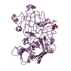
| ||||||||
|---|---|---|---|---|---|---|---|---|---|
| 1 |
| ||||||||
| Unit cell |
| ||||||||
| Atom site foot note | 1: RESIDUES PRO E 134 AND PRO E 315 ARE CIS PROLINES. 2: THE REGION FROM SER E 277 TO SER E 281 IS POORLY ORDERED. 3: WATER MOLECULE 411, IS SITTING ON A SPECIAL POSITION. | ||||||||
| Components on special symmetry positions |
|
-Components
| #1: Protein | Mass: 33468.809 Da / Num. of mol.: 1 Source method: isolated from a genetically manipulated source Source: (gene. exp.) Penicillium janthinellum (fungus) / References: UniProt: P00798, penicillopepsin |
|---|---|
| #2: Protein/peptide | Mass: 500.671 Da / Num. of mol.: 1 Source method: isolated from a genetically manipulated source Details: TRANSITION STATE MIMIC INHIBITOR |
| #3: Sugar | ChemComp-MAN /   Type: D-saccharide, alpha linking / Mass: 180.156 Da / Num. of mol.: 1 Type: D-saccharide, alpha linking / Mass: 180.156 Da / Num. of mol.: 1Source method: isolated from a genetically manipulated source Formula: C6H12O6 |
| #4: Water | ChemComp-HOH /  Mass: 18.015 Da / Num. of mol.: 247 / Source method: isolated from a natural source / Formula: H2O Mass: 18.015 Da / Num. of mol.: 247 / Source method: isolated from a natural source / Formula: H2O |
| Has protein modification | Y |
| Nonpolymer details | LTA IS AN O-ETHYL ANALOGUE OF STATINE IN WHICH THE LEUCINE-LIKE SIDE CHAIN OF STATINE HAS BEEN ...LTA IS AN O-ETHYL ANALOGUE OF STATINE IN WHICH THE LEUCINE-LIKE SIDE CHAIN OF STATINE HAS BEEN REPLACED BY THE LYSINE-LIKE SIDE CHAIN. |
-Experimental details
-Experiment
| Experiment | Method: X-RAY DIFFRACTION |
|---|
- Sample preparation
| Crystal | Density Matthews: 1.99 Å3/Da / Density % sol: 38.12 % |
|---|
-Data collection
| Radiation | Scattering type: x-ray |
|---|---|
| Radiation wavelength | Relative weight: 1 |
- Processing
| Software | Name: PROLSQ / Classification: refinement | |||||||||||||||||||||||||||||||||||||||||||||||||||||||||||||||
|---|---|---|---|---|---|---|---|---|---|---|---|---|---|---|---|---|---|---|---|---|---|---|---|---|---|---|---|---|---|---|---|---|---|---|---|---|---|---|---|---|---|---|---|---|---|---|---|---|---|---|---|---|---|---|---|---|---|---|---|---|---|---|---|---|
| Refinement | Resolution: 1.8→8 Å / σ(I): 1 /
| |||||||||||||||||||||||||||||||||||||||||||||||||||||||||||||||
| Refinement step | Cycle: LAST / Resolution: 1.8→8 Å
| |||||||||||||||||||||||||||||||||||||||||||||||||||||||||||||||
| Refine LS restraints |
|
