 Movie
Movie Controller

[English] 日本語
 Yorodumi
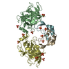
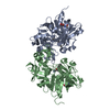
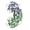
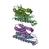
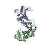
- PDB-2web: ACID PROTEINASE (PENICILLOPEPSIN) (E.C.3.4.23.20) COMPLEX WITH PH... -
+ Open data

- Basic information
| Entry | Database: PDB / ID: 2web | |||||||||
|---|---|---|---|---|---|---|---|---|---|---|
| Title | ACID PROTEINASE (PENICILLOPEPSIN) (E.C.3.4.23.20) COMPLEX WITH PHOSPHONATE INHIBITOR: METHYL(2S)-[1-(((N-FORMYL)-L-VALYL)AMINO-2-(2-NAPHTHYL)ETHYL)HYDROXYPHOSPHINYLOXY]-3-PHENYLPROPANOATE, SODIUM SALT | |||||||||
 Components Components | PENICILLOPEPSIN | |||||||||
 Keywords Keywords | HYDROLASE / PENICILLOPEPSIN / PHOSPHONATE INHIBITOR | |||||||||
| Function / homology |  Function and homology information Function and homology informationpenicillopepsin / aspartic-type endopeptidase activity / proteolysis / extracellular region Similarity search - Function | |||||||||
| Biological species |  Penicillium janthinellum (fungus) Penicillium janthinellum (fungus) | |||||||||
| Method |  X-RAY DIFFRACTION / DIFFERENCE FOURIER METHOD / Resolution: 1.5 Å X-RAY DIFFRACTION / DIFFERENCE FOURIER METHOD / Resolution: 1.5 Å | |||||||||
 Authors Authors | Ding, J. / Fraser, M.E. / James, M.N.G. | |||||||||
 Citation Citation | Journal: J.Am.Chem.Soc. / Year: 1998 Title: Macrocyclic Inhibitors of Penicillopepsin. II. X-Ray Crystallographic Analyses of Penicillopepsin Complexed with a P3-P1 Macrocyclic Peptidyl Inhibitor and with its Two Acyclic Analogues Authors: Ding, J. / Fraser, M.E. / Meyer, J.H. / Bartlett, P.A. / James, M.N.G. #1: Journal: To be PublishedTitle: Macrocyclic Inhibitors of Penicillopepsin. I. Design, Synthesis, and Evaluation of an Inhibitor Bridged between P1 and P3 Authors: Meyer, J.H. / Bartlett, P.A. #2: Journal: Biochemistry / Year: 1992Title: Crystallographic Analysis of Transition-State Mimics Bound to Penicillopepsin: Phosphorus-Containing Peptide Analogues Authors: Fraser, M.E. / Strynadka, N.C. / Bartlett, P.A. / Hanson, J.E. / James, M.N. #3: Journal: Biochemistry / Year: 1992Title: Crystallographic Analysis of Transition State Mimics Bound to Penicillopepsin: Difluorostatine-and Difluorostatone-Containing Peptides Authors: James, M.N. / Sielecki, A.R. / Hayakawa, K. / Gelb, M.H. | |||||||||
| History |
|
- Structure visualization
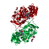
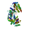
| Structure viewer | Molecule: MolmilJmol/JSmol |
|---|
- Downloads & links
-Download
| PDBx/mmCIF format | 2web.cif.gz | 81.8 KB | Display | PDBx/mmCIF format |
|---|---|---|---|---|
| PDB format | pdb2web.ent.gz | 59.6 KB | Display | PDB format |
| PDBx/mmJSON format | 2web.json.gz | Tree view | PDBx/mmJSON format | |
| Others |  Other downloads Other downloads |
-Validation report
| Arichive directory | https://data.pdbj.org/pub/pdb/validation_reports/we/2webftp://data.pdbj.org/pub/pdb/validation_reports/we/2web | HTTPS FTP |
|---|
-Related structure data
| Similar structure data |
|---|


-Links
 PDBj
PDBj


- Assembly
| Deposited unit | 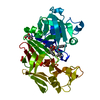
| ||||||||
|---|---|---|---|---|---|---|---|---|---|
| 1 | 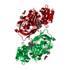
| ||||||||
| Unit cell |
|
-Components
| #1: Protein | Mass: 33468.809 Da / Num. of mol.: 1 / Source method: isolated from a natural source / Source: (natural) Penicillium janthinellum (fungus) / References: UniProt: P00798, penicillopepsin | ||||||||
|---|---|---|---|---|---|---|---|---|---|
| #2: Sugar |   Type: D-saccharide, alpha linking / Mass: 180.156 Da / Num. of mol.: 2 Type: D-saccharide, alpha linking / Mass: 180.156 Da / Num. of mol.: 2Source method: isolated from a genetically manipulated source Formula: C6H12O6 #3: Chemical | ChemComp-SO4 / |   Mass: 96.063 Da / Num. of mol.: 1 / Source method: obtained synthetically / Formula: SO4 Mass: 96.063 Da / Num. of mol.: 1 / Source method: obtained synthetically / Formula: SO4#4: Chemical | ChemComp-PP4 / |   Mass: 539.537 Da / Num. of mol.: 1 / Source method: obtained synthetically / Formula: C28H32N2O7P Mass: 539.537 Da / Num. of mol.: 1 / Source method: obtained synthetically / Formula: C28H32N2O7P#5: Water | ChemComp-HOH / |  Mass: 18.015 Da / Num. of mol.: 323 / Source method: isolated from a natural source / Formula: H2O Mass: 18.015 Da / Num. of mol.: 323 / Source method: isolated from a natural source / Formula: H2OHas protein modification | Y | |
-Experimental details
-Experiment
| Experiment | Method: X-RAY DIFFRACTION / Number of used crystals: 1 |
|---|
- Sample preparation
| Crystal | Density Matthews: 2.02 Å3/Da / Density % sol: 39.03 % |
|---|---|
| Crystal grow | pH: 4.4 / Details: 0.1M NAC2H3O2 PH=4.4 35-40% SATURATED (NH4)2SO4 |
-Data collection
| Diffraction | Mean temperature: 293 K |
|---|---|
| Diffraction source | Source: ROTATING ANODE / Type: OTHER / Wavelength: 1.5418 |
| Detector | Type: SIEMENS-NICOLET X100 / Detector: AREA DETECTOR / Date: Apr 25, 1997 |
| Radiation | Monochromator: GRAPHITE(002) / Monochromatic (M) / Laue (L): M / Scattering type: x-ray |
| Radiation wavelength | Wavelength: 1.5418 Å / Relative weight: 1 |
| Reflection | Resolution: 1.5→40 Å / Num. obs: 40665 / % possible obs: 94.74 % / Observed criterion σ(I): 0 / Redundancy: 5.76 % / Biso Wilson estimate: 14.946 Å2 / Rmerge(I) obs: 0.0836 / Net I/σ(I): 33.83 |
| Reflection shell | Resolution: 1.5→1.55 Å / Redundancy: 3.09 % / Rmerge(I) obs: 0.272 / Mean I/σ(I) obs: 2.886 / % possible all: 78.94 |
- Processing
| Software |
| ||||||||||||||||||||||||||||||||||||||||||||||||||
|---|---|---|---|---|---|---|---|---|---|---|---|---|---|---|---|---|---|---|---|---|---|---|---|---|---|---|---|---|---|---|---|---|---|---|---|---|---|---|---|---|---|---|---|---|---|---|---|---|---|---|---|
| Refinement | Method to determine structure: DIFFERENCE FOURIER METHOD / Resolution: 1.5→40 Å Isotropic thermal model: CSDX_PROTGEO.DAT, OTHERGEO.DAT, PP4GEO.DAT, SEMTHMGEO.DAT σ(F): 2 Stereochemistry target values: CSDX_PROTGEO.DAT, OTHERGEO.DAT, PP4GEO.DAT, SEMTHMGEO.DAT, CONTACT.DAT Details: X-PLOR AND TNT ESD FROM SIGMAA (A) : 0.191880 UNCERTAINTY IN RMS ERROR SQUARED : 0.005343
| ||||||||||||||||||||||||||||||||||||||||||||||||||
| Solvent computation | Bsol: 357.8 Å2 / ksol: 0.851 e/Å3 | ||||||||||||||||||||||||||||||||||||||||||||||||||
| Refinement step | Cycle: LAST / Resolution: 1.5→40 Å
| ||||||||||||||||||||||||||||||||||||||||||||||||||
| Refine LS restraints |
|
