 Movie
Movie Controller

[English] 日本語
 Yorodumi
- PDB-2r9p: Human mesotrypsin complexed with bovine pancreatic trypsin inhibi... -
+ Open data

- Basic information
| Entry | Database: PDB / ID: 2r9p | ||||||
|---|---|---|---|---|---|---|---|
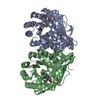
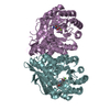
| Title | Human mesotrypsin complexed with bovine pancreatic trypsin inhibitor(BPTI) | ||||||
 Components Components |
| ||||||
 Keywords Keywords | hydrolase/hydrolase inhibitor / Human mesotrypsin / Serine protease / Bovine pancreatic trypsin inhibitor / BPTI / Alternative splicing / Calcium / Digestion / Hydrolase / Metal-binding / Secreted / Sulfation / Zymogen / Pharmaceutical / Protease inhibitor / Serine protease inhibitor / hydrolase-hydrolase inhibitor COMPLEX | ||||||
| Function / homology |  Function and homology information Function and homology informationUptake of dietary cobalamins into enterocytes / sulfate binding / negative regulation of platelet aggregation / zymogen binding / antimicrobial humoral response / Alpha-defensins / potassium channel inhibitor activity / Differentiation of Keratinocytes in Interfollicular Epidermis in Mammalian Skin / Antimicrobial peptides / zymogen activation ...Uptake of dietary cobalamins into enterocytes / sulfate binding / negative regulation of platelet aggregation / zymogen binding / antimicrobial humoral response / Alpha-defensins / potassium channel inhibitor activity / Differentiation of Keratinocytes in Interfollicular Epidermis in Mammalian Skin / Antimicrobial peptides / zymogen activation / molecular function inhibitor activity / negative regulation of thrombin-activated receptor signaling pathway / trypsin / endothelial cell migration / serine protease inhibitor complex / digestion / serine-type peptidase activity / serine-type endopeptidase inhibitor activity / tertiary granule lumen / protease binding / serine-type endopeptidase activity / calcium ion binding / Neutrophil degranulation / proteolysis / : / extracellular region Similarity search - Function | ||||||
| Biological species |  Homo sapiens (human) Homo sapiens (human) | ||||||
| Method |  X-RAY DIFFRACTION / SYNCHROTRON / MOLECULAR REPLACEMENT / Resolution: 1.4 Å X-RAY DIFFRACTION / SYNCHROTRON / MOLECULAR REPLACEMENT / Resolution: 1.4 Å | ||||||
 Authors Authors | Salameh, M.A. / Soares, A.S. / Radisky, E.S. | ||||||
 Citation Citation | Journal: J.Biol.Chem. / Year: 2008 Title: Structural Basis for Accelerated Cleavage of Bovine Pancreatic Trypsin Inhibitor (BPTI) by Human Mesotrypsin. Authors: Salameh, M.A. / Soares, A.S. / Hockla, A. / Radisky, E.S. | ||||||
| History |
|
- Structure visualization
| Structure viewer | Molecule: MolmilJmol/JSmol |
|---|
- Downloads & links
-Download
| PDBx/mmCIF format | 2r9p.cif.gz | 241.5 KB | Display | PDBx/mmCIF format |
|---|---|---|---|---|
| PDB format | pdb2r9p.ent.gz | 193.5 KB | Display | PDB format |
| PDBx/mmJSON format | 2r9p.json.gz | Tree view | PDBx/mmJSON format | |
| Others |  Other downloads Other downloads |
-Validation report
| Arichive directory | https://data.pdbj.org/pub/pdb/validation_reports/r9/2r9pftp://data.pdbj.org/pub/pdb/validation_reports/r9/2r9p | HTTPS FTP |
|---|
-Related structure data
| Related structure data |  2ra3C 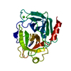 1h4wS 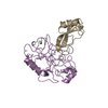 2ptcS C: citing same article ( S: Starting model for refinement |
|---|---|
| Similar structure data |


-Links
 PDBj
PDBj


- Assembly
| Deposited unit | 
| ||||||||
|---|---|---|---|---|---|---|---|---|---|
| 1 | 
| ||||||||
| 2 | 
| ||||||||
| Unit cell |
|
-Components
| #1: Protein | Mass: 24257.457 Da / Num. of mol.: 4 / Mutation: S195A Source method: isolated from a genetically manipulated source Source: (gene. exp.) Homo sapiens (human) / Gene: PRSS3, PRSS4, TRY3, TRY4 / Production host:  #2: Protein | Mass: 6527.568 Da / Num. of mol.: 4 / Source method: isolated from a natural source / Source: (natural) #3: Chemical | ChemComp-SO4 /   Mass: 96.063 Da / Num. of mol.: 20 / Source method: obtained synthetically / Formula: SO4 Mass: 96.063 Da / Num. of mol.: 20 / Source method: obtained synthetically / Formula: SO4#4: Water | ChemComp-HOH / |  Mass: 18.015 Da / Num. of mol.: 633 / Source method: isolated from a natural source / Formula: H2O Mass: 18.015 Da / Num. of mol.: 633 / Source method: isolated from a natural source / Formula: H2OHas protein modification | Y | |
|---|
-Experimental details
-Experiment
| Experiment | Method: X-RAY DIFFRACTION / Number of used crystals: 1 |
|---|
- Sample preparation
| Crystal | Density Matthews: 2.39 Å3/Da / Density % sol: 48.5 % |
|---|---|
| Crystal grow | Temperature: 298 K / Method: vapor diffusion, hanging drop / pH: 5.3 Details: 1.6M ammonium sulfate, pH 5.3, VAPOR DIFFUSION, HANGING DROP, temperature 298.0K |
-Data collection
| Diffraction | Mean temperature: 100 K |
|---|---|
| Diffraction source | Source: SYNCHROTRON / Site: NSLS  / Beamline: X12B / Wavelength: 1.1 Å / Beamline: X12B / Wavelength: 1.1 Å |
| Detector | Type: ADSC QUANTUM 4 / Detector: CCD / Date: Apr 25, 2007 |
| Radiation | Monochromator: Si 111 CHANNEL / Protocol: SINGLE WAVELENGTH / Monochromatic (M) / Laue (L): M / Scattering type: x-ray |
| Radiation wavelength | Wavelength: 1.1 Å / Relative weight: 1 |
| Reflection | Resolution: 1.4→50 Å / Num. obs: 221478 / % possible obs: 97.8 % / Observed criterion σ(F): -3 / Observed criterion σ(I): -3 / Redundancy: 4 % / Biso Wilson estimate: 18.803 Å2 / Rmerge(I) obs: 0.081 / Rsym value: 0.081 / Net I/σ(I): 15.275 |
| Reflection shell | Resolution: 1.4→1.45 Å / Redundancy: 3.3 % / Rmerge(I) obs: 0.84 / Mean I/σ(I) obs: 3.2 / Rsym value: 0.84 / % possible all: 96.2 |
- Processing
| Software |
| |||||||||||||||||||||||||
|---|---|---|---|---|---|---|---|---|---|---|---|---|---|---|---|---|---|---|---|---|---|---|---|---|---|---|
| Refinement | Method to determine structure: MOLECULAR REPLACEMENT Starting model: PDB entries 1H4W and 2PTC Resolution: 1.4→25.6 Å / Cross valid method: THROUGHOUT / σ(F): 0 / σ(I): 0 / Stereochemistry target values: Engh & Huber
| |||||||||||||||||||||||||
| Displacement parameters | Biso mean: 18.8 Å2 | |||||||||||||||||||||||||
| Refinement step | Cycle: LAST / Resolution: 1.4→25.6 Å
| |||||||||||||||||||||||||
| Refine LS restraints |
| |||||||||||||||||||||||||
| LS refinement shell | Resolution: 1.4→1.45 Å |
