[English] 日本語
Yorodumi
- PDB-4xhr: Structure of a phospholipid trafficking complex, native -

+
Open data


ID or keywords:

Loading...

-
Basic information

Entry
Database: PDB / ID: 4xhr
TitleStructure of a phospholipid trafficking complex, native
Components
  • Mitochondrial distribution and morphology protein 35
  • Protein UPS1, mitochondrial
KeywordsLIPID TRANSPORT/OXIDOREDUCTASE / phospholipid / LIPID TRANSPORT-OXIDOREDUCTASE complex
Function / homology
Function and homology information


TP53 Regulates Transcription of Genes Involved in Cytochrome C Release / phosphatidic acid transfer activity / cardiolipin metabolic process / positive regulation of phosphatidylcholine biosynthetic process / mitochondrial respiratory chain complex assembly / intermembrane lipid transfer / phospholipid transport / phospholipid translocation / mitochondrion organization / mitochondrial intermembrane space ...TP53 Regulates Transcription of Genes Involved in Cytochrome C Release / phosphatidic acid transfer activity / cardiolipin metabolic process / positive regulation of phosphatidylcholine biosynthetic process / mitochondrial respiratory chain complex assembly / intermembrane lipid transfer / phospholipid transport / phospholipid translocation / mitochondrion organization / mitochondrial intermembrane space / mitochondrial inner membrane / lipid binding / mitochondrion / nucleus / cytoplasm
Similarity search - Function
PRELI/MSF1 domain / Slowmo/Ups family / PRELI-like family / PRELI/MSF1 domain profile. / Mitochondrial distribution/morphology family 35/apoptosis / Uncharacterised protein family (UPF0203) / Coiled coil-helix-coiled coil-helix (CHCH) domain profile.
Similarity search - Domain/homology
Mitochondrial distribution and morphology protein 35 / Protein UPS1, mitochondrial
Similarity search - Component
Biological speciesSaccharomyces cerevisiae (brewer's yeast)
MethodX-RAY DIFFRACTION / SYNCHROTRON / SAD / Resolution: 2.55 Å
AuthorsYu, F. / He, F. / Wang, C. / Zhang, P.
CitationJournal: Embo Rep. / Year: 2015
Title: Structural basis of intramitochondrial phosphatidic acid transport mediated by Ups1-Mdm35 complex
Authors: Yu, F. / He, F. / Yao, H. / Wang, C. / Wang, J. / Li, J. / Qi, X. / Xue, H. / Ding, J. / Zhang, P.
History
DepositionJan 6, 2015Deposition site: RCSB / Processing site: PDBJ
Revision 1.0Jul 1, 2015Provider: repository / Type: Initial release
Revision 1.1Aug 5, 2015Group: Database references

-
Structure visualization

Structure viewerMolecule:
MolmilJmol/JSmol

Downloads & links

-
Assembly

Deposited unit
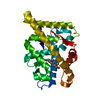
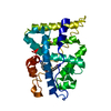
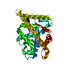
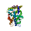
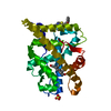
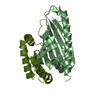
A: Protein UPS1, mitochondrial
N: Mitochondrial distribution and morphology protein 35
B: Protein UPS1, mitochondrial
M: Mitochondrial distribution and morphology protein 35


Theoretical massNumber of molelcules
Total (without water)62,9594
Polymers62,9594
Non-polymers00
Water68538
1
A: Protein UPS1, mitochondrial
N: Mitochondrial distribution and morphology protein 35


Theoretical massNumber of molelcules
Total (without water)31,4802
Polymers31,4802
Non-polymers00
Water362
TypeNameSymmetry operationNumber
identity operation1_555x,y,z1
Buried area2740 Å2
ΔGint-17 kcal/mol
Surface area12560 Å2
MethodPISA
2
B: Protein UPS1, mitochondrial
M: Mitochondrial distribution and morphology protein 35


Theoretical massNumber of molelcules
Total (without water)31,4802
Polymers31,4802
Non-polymers00
Water362
TypeNameSymmetry operationNumber
identity operation1_555x,y,z1
Buried area2700 Å2
ΔGint-18 kcal/mol
Surface area13090 Å2
MethodPISA
Unit cell
Length a, b, c (Å)42.968, 74.868, 87.568
Angle α, β, γ (deg.)90.00, 95.32, 90.00
Int Tables number4
Space group name H-MP1211

-
Components

#1: Protein Protein UPS1, mitochondrial / Unprocessed MGM1 protein 1


Mass: 21755.670 Da / Num. of mol.: 2
Source method: isolated from a genetically manipulated source
Source: (gene. exp.) Saccharomyces cerevisiae (strain ATCC 204508 / S288c) (yeast)
Strain: ATCC 204508 / S288c / Gene: UPS1, YLR193C / Production host: Escherichia coli (E. coli) / References: UniProt: Q05776
#2: Protein Mitochondrial distribution and morphology protein 35


Mass: 9723.954 Da / Num. of mol.: 2
Source method: isolated from a genetically manipulated source
Source: (gene. exp.) Saccharomyces cerevisiae (strain ATCC 204508 / S288c) (yeast)
Strain: ATCC 204508 / S288c / Gene: MDM35, YKL053C-A / Production host: Escherichia coli (E. coli) / References: UniProt: O60200
#3: Water ChemComp-HOH / water / Water


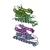
Mass: 18.015 Da / Num. of mol.: 38 / Source method: isolated from a natural source / Formula: H2O

-
Experimental details

-
Experiment

ExperimentMethod: X-RAY DIFFRACTION

-
Sample preparation

CrystalDensity Matthews: 2.23 Å3/Da / Density % sol: 44.78 %
Crystal growTemperature: 277.15 K / Method: vapor diffusion, sitting drop
Details: 2%(v/v) Tacsimate pH 6.0, 0.1M Bis-Tris pH 6.5, 20%(w/v) PEG3350

-
Data collection

DiffractionMean temperature: 100 K
Diffraction sourceSource: SYNCHROTRON / Site: SSRF / Beamline: BL17U / Wavelength: 1.7 Å
DetectorType: MARMOSAIC 225 mm CCD / Detector: CCD / Date: Jul 8, 2013
RadiationProtocol: SINGLE WAVELENGTH / Monochromatic (M) / Laue (L): M / Scattering type: x-ray
Radiation wavelengthWavelength: 1.7 Å / Relative weight: 1
ReflectionResolution: 2.55→32.08 Å / Num. obs: 17640 / % possible obs: 97.17 % / Redundancy: 3.5 % / Net I/σ(I): 11.96
Reflection shellResolution: 2.55→3 Å / Redundancy: 3.5 % / Rmerge(I) obs: 0.116 / Mean I/σ(I) obs: 11.96 / % possible all: 97.17

-
Processing

Software
NameVersionClassification
REFMAC5.7.0029refinement
HKL-2000data reduction
HKL-2000data scaling
PHASERphasing
RefinementMethod to determine structure: SAD / Resolution: 2.55→32.08 Å / Cor.coef. Fo:Fc: 0.924 / Cor.coef. Fo:Fc free: 0.891 / SU B: 25.837 / SU ML: 0.26 / Cross valid method: THROUGHOUT / ESU R: 1.232 / ESU R Free: 0.353 / Stereochemistry target values: MAXIMUM LIKELIHOOD / Details: HYDROGENS HAVE BEEN ADDED IN THE RIDING POSITIONS
RfactorNum. reflection% reflectionSelection details
Rfree0.27736 886 5.1 %RANDOM
Rwork0.2363 ---
obs0.23837 16623 96.73 %-
Solvent computationIon probe radii: 0.8 Å / Shrinkage radii: 0.8 Å / VDW probe radii: 1.2 Å / Solvent model: MASK
Displacement parametersBiso mean: 78.621 Å2
Baniso -1Baniso -2Baniso -3
1--5.72 Å20 Å22.49 Å2
2--6.37 Å20 Å2
3----0.87 Å2
Refinement stepCycle: LAST / Resolution: 2.55→32.08 Å
ProteinNucleic acidLigandSolventTotal
Num. atoms3869 0 0 38 3907
Refine LS restraints
Refine-IDTypeDev idealDev ideal targetNumber
X-RAY DIFFRACTIONr_bond_refined_d0.150.023961
X-RAY DIFFRACTIONr_bond_other_d00.023726
X-RAY DIFFRACTIONr_angle_refined_deg1.8611.9435350
X-RAY DIFFRACTIONr_angle_other_deg3.5083.0028601
X-RAY DIFFRACTIONr_dihedral_angle_1_deg7.9995476
X-RAY DIFFRACTIONr_dihedral_angle_2_deg36.93623.864176
X-RAY DIFFRACTIONr_dihedral_angle_3_deg18.48615708
X-RAY DIFFRACTIONr_dihedral_angle_4_deg18.6791522
X-RAY DIFFRACTIONr_chiral_restr0.070.2587
X-RAY DIFFRACTIONr_gen_planes_refined0.0090.0214378
X-RAY DIFFRACTIONr_gen_planes_other0.0050.02916
X-RAY DIFFRACTIONr_nbd_refined
X-RAY DIFFRACTIONr_nbd_other
X-RAY DIFFRACTIONr_nbtor_refined
X-RAY DIFFRACTIONr_nbtor_other
X-RAY DIFFRACTIONr_xyhbond_nbd_refined
X-RAY DIFFRACTIONr_xyhbond_nbd_other
X-RAY DIFFRACTIONr_metal_ion_refined
X-RAY DIFFRACTIONr_metal_ion_other
X-RAY DIFFRACTIONr_symmetry_vdw_refined
X-RAY DIFFRACTIONr_symmetry_vdw_other
X-RAY DIFFRACTIONr_symmetry_hbond_refined
X-RAY DIFFRACTIONr_symmetry_hbond_other
X-RAY DIFFRACTIONr_symmetry_metal_ion_refined
X-RAY DIFFRACTIONr_symmetry_metal_ion_other
X-RAY DIFFRACTIONr_mcbond_it
X-RAY DIFFRACTIONr_mcbond_other
X-RAY DIFFRACTIONr_mcangle_it
X-RAY DIFFRACTIONr_mcangle_other
X-RAY DIFFRACTIONr_scbond_it
X-RAY DIFFRACTIONr_scbond_other
X-RAY DIFFRACTIONr_scangle_it
X-RAY DIFFRACTIONr_scangle_other
X-RAY DIFFRACTIONr_long_range_B_refined
X-RAY DIFFRACTIONr_long_range_B_other
X-RAY DIFFRACTIONr_rigid_bond_restr
X-RAY DIFFRACTIONr_sphericity_free
X-RAY DIFFRACTIONr_sphericity_bonded
LS refinement shellResolution: 2.55→2.619 Å / Total num. of bins used: 20
RfactorNum. reflection% reflection
Rfree0.374 52 -
Rwork0.314 1250 -
obs--97.38 %
Refinement TLS params.

Method: refined / Refine-ID: X-RAY DIFFRACTION

IDL112)L122)L132)L222)L232)L332)S11 (Å °)S12 (Å °)S13 (Å °)S21 (Å °)S22 (Å °)S23 (Å °)S31 (Å °)S32 (Å °)S33 (Å °)T112)T122)T132)T222)T232)T332)Origin x (Å)Origin y (Å)Origin z (Å)
11.24480.3320.24340.55320.35691.89540.028-0.19970.0061-0.0433-0.01690.046-0.07640.089-0.01110.1489-0.0032-0.07430.0526-0.03440.126560.4324-10.7839.2214
21.65050.1899-0.55970.93040.51822.50060.00580.323-0.336-0.07830.08430.0344-0.04040.0466-0.09010.2133-0.0026-0.07840.08-0.06870.143760.1617-14.079220.7976
31.85360.02440.04730.53270.56111.8234-0.0187-0.0128-0.02670.0155-0.0173-0.0166-0.08560.06770.0360.13350.0177-0.09590.0161-0.00050.11260.6055-42.979716.6653
44.57080.78870.08441.5161-0.2782.1093-0.04130.59350.12070.01630.08330.1612-0.18740.1155-0.0420.19330.0396-0.06580.2393-0.03060.047760.4373-40.9843-1.8287
Refinement TLS group
IDRefine-IDRefine TLS-IDAuth asym-IDAuth seq-ID
1X-RAY DIFFRACTION1A1 - 170
2X-RAY DIFFRACTION2N6 - 76
3X-RAY DIFFRACTION3B1 - 170
4X-RAY DIFFRACTION4M5 - 76

+
About Yorodumi

-
News

-
Feb 9, 2022. New format data for meta-information of EMDB entries

New format data for meta-information of EMDB entries

  • Version 3 of the EMDB header file is now the official format.
  • The previous official version 1.9 will be removed from the archive.

Related info.:EMDB header

External links:wwPDB to switch to version 3 of the EMDB data model

-
Aug 12, 2020. Covid-19 info

Covid-19 info

URL: https://pdbj.org/emnavi/covid19.php

New page: Covid-19 featured information page in EM Navigator.

Related info.:Covid-19 info / Mar 5, 2020. Novel coronavirus structure data

+
Mar 5, 2020. Novel coronavirus structure data

Novel coronavirus structure data

Related info.:Yorodumi Speices / Aug 12, 2020. Covid-19 info

External links:COVID-19 featured content - PDBj / Molecule of the Month (242):Coronavirus Proteases

+
Jan 31, 2019. EMDB accession codes are about to change! (news from PDBe EMDB page)

EMDB accession codes are about to change! (news from PDBe EMDB page)

  • The allocation of 4 digits for EMDB accession codes will soon come to an end. Whilst these codes will remain in use, new EMDB accession codes will include an additional digit and will expand incrementally as the available range of codes is exhausted. The current 4-digit format prefixed with “EMD-” (i.e. EMD-XXXX) will advance to a 5-digit format (i.e. EMD-XXXXX), and so on. It is currently estimated that the 4-digit codes will be depleted around Spring 2019, at which point the 5-digit format will come into force.
  • The EM Navigator/Yorodumi systems omit the EMD- prefix.

Related info.:Q: What is EMD? / ID/Accession-code notation in Yorodumi/EM Navigator

External links:EMDB Accession Codes are Changing Soon! / Contact to PDBj

+
Jul 12, 2017. Major update of PDB

Major update of PDB

  • wwPDB released updated PDB data conforming to the new PDBx/mmCIF dictionary.
  • This is a major update changing the version number from 4 to 5, and with Remediation, in which all the entries are updated.
  • In this update, many items about electron microscopy experimental information are reorganized (e.g. em_software).
  • Now, EM Navigator and Yorodumi are based on the updated data.

External links:wwPDB Remediation / Enriched Model Files Conforming to OneDep Data Standards Now Available in the PDB FTP Archive

-
Yorodumi

Thousand views of thousand structures

  • Yorodumi is a browser for structure data from EMDB, PDB, SASBDB, etc.
  • This page is also the successor to EM Navigator detail page, and also detail information page/front-end page for Omokage search.
  • The word "yorodu" (or yorozu) is an old Japanese word meaning "ten thousand". "mi" (miru) is to see.

Related info.:EMDB / PDB / SASBDB / Comparison of 3 databanks / Yorodumi Search / Aug 31, 2016. New EM Navigator & Yorodumi / Yorodumi Papers / Jmol/JSmol / Function and homology information / Changes in new EM Navigator and Yorodumi

Read more