 Movie
Movie Controller

[English] 日本語
 Yorodumi
- PDB-4ua7: CTX-M-14 Class A Beta-Lactamase in Complex with a Non-Covalent In... -
+ Open data

- Basic information
| Entry | Database: PDB / ID: 4ua7 | |||||||||
|---|---|---|---|---|---|---|---|---|---|---|
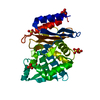
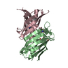
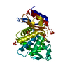
| Title | CTX-M-14 Class A Beta-Lactamase in Complex with a Non-Covalent Inhibitor at Sub-Angstrom Resolution | |||||||||
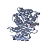
 Components Components | Beta-lactamase CTX-M-14 | |||||||||
 Keywords Keywords | HYDROLASE/HYDROLASE Inhibitor / CTX-M-14 / Class A Beta-Lactamase / Ultra High Resolution / HYDROLASE-HYDROLASE Inhibitor complex | |||||||||
| Function / homology |  Function and homology information Function and homology informationbeta-lactam antibiotic catabolic process / beta-lactamase activity / beta-lactamase / response to antibiotic Similarity search - Function | |||||||||
| Biological species |  | |||||||||
| Method |  X-RAY DIFFRACTION / SYNCHROTRON / MOLECULAR REPLACEMENT / Resolution: 0.89 Å X-RAY DIFFRACTION / SYNCHROTRON / MOLECULAR REPLACEMENT / Resolution: 0.89 Å | |||||||||
 Authors Authors | Nichols, D.A. / Chen, Y. | |||||||||
| Funding support |  United States, 1items United States, 1items
| |||||||||
 Citation Citation | Journal: J.Am.Chem.Soc. / Year: 2015 Title: Ligand-Induced Proton Transfer and Low-Barrier Hydrogen Bond Revealed by X-ray Crystallography. Authors: Nichols, D.A. / Hargis, J.C. / Sanishvili, R. / Jaishankar, P. / Defrees, K. / Smith, E.W. / Wang, K.K. / Prati, F. / Renslo, A.R. / Woodcock, H.L. / Chen, Y. | |||||||||
| History |
|
- Structure visualization
| Structure viewer | Molecule: MolmilJmol/JSmol |
|---|
- Downloads & links
-Download
| PDBx/mmCIF format | 4ua7.cif.gz | 369 KB | Display | PDBx/mmCIF format |
|---|---|---|---|---|
| PDB format | pdb4ua7.ent.gz | 307.4 KB | Display | PDB format |
| PDBx/mmJSON format | 4ua7.json.gz | Tree view | PDBx/mmJSON format | |
| Others |  Other downloads Other downloads |
-Validation report
| Arichive directory | https://data.pdbj.org/pub/pdb/validation_reports/ua/4ua7ftp://data.pdbj.org/pub/pdb/validation_reports/ua/4ua7 | HTTPS FTP |
|---|
-Related structure data
| Related structure data |  4ua6C  4ua9C  4uaaC  2p74S C: citing same article ( S: Starting model for refinement |
|---|---|
| Similar structure data |


-Links
 PDBj
PDBj


- Assembly
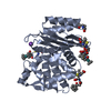
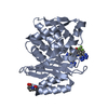
| Deposited unit | 
| ||||||||
|---|---|---|---|---|---|---|---|---|---|
| 1 | 
| ||||||||
| 2 | 
| ||||||||
| Unit cell |
|
-Components
| #1: Protein | Mass: 27983.516 Da / Num. of mol.: 2 Source method: isolated from a genetically manipulated source Source: (gene. exp.) #2: Chemical | ChemComp-3GK / 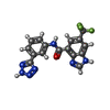  Mass: 373.292 Da / Num. of mol.: 4 / Source method: obtained synthetically / Formula: C16H10F3N7O Mass: 373.292 Da / Num. of mol.: 4 / Source method: obtained synthetically / Formula: C16H10F3N7O#3: Chemical |   Mass: 94.971 Da / Num. of mol.: 3 / Source method: obtained synthetically / Formula: PO4 Mass: 94.971 Da / Num. of mol.: 3 / Source method: obtained synthetically / Formula: PO4#4: Water | ChemComp-HOH / |  Mass: 18.015 Da / Num. of mol.: 870 / Source method: isolated from a natural source / Formula: H2O Mass: 18.015 Da / Num. of mol.: 870 / Source method: isolated from a natural source / Formula: H2OHas protein modification | Y | |
|---|
-Experimental details
-Experiment
| Experiment | Method: X-RAY DIFFRACTION / Number of used crystals: 1 |
|---|
- Sample preparation
| Crystal | Density Matthews: 2.01 Å3/Da / Density % sol: 38.8 % |
|---|---|
| Crystal grow | Temperature: 293 K / Method: vapor diffusion, hanging drop / pH: 8.3 / Details: 1.0M Potassium Phosphate / PH range: 8.3 |
-Data collection
| Diffraction | Mean temperature: 100 K |
|---|---|
| Diffraction source | Source: SYNCHROTRON / Site: ALS / Beamline: 8.3.1 / Wavelength: 0.7749 Å |
| Detector | Type: ADSC QUANTUM 315r / Detector: CCD / Date: Aug 7, 2012 |
| Radiation | Protocol: SINGLE WAVELENGTH / Monochromatic (M) / Laue (L): M / Scattering type: x-ray |
| Radiation wavelength | Wavelength: 0.7749 Å / Relative weight: 1 |
| Reflection | Resolution: 0.89→50 Å / Num. obs: 334913 / % possible obs: 99.4 % / Redundancy: 4 % / Rmerge(I) obs: 0.052 / Net I/σ(I): 22 |
- Processing
| Software |
| |||||||||||||||||||||||||||||||||
|---|---|---|---|---|---|---|---|---|---|---|---|---|---|---|---|---|---|---|---|---|---|---|---|---|---|---|---|---|---|---|---|---|---|---|
| Refinement | Method to determine structure: MOLECULAR REPLACEMENT Starting model: 2P74 Resolution: 0.89→10 Å / Num. parameters: 50213 / Num. restraintsaints: 68566 / Cross valid method: FREE R-VALUE / σ(F): 0 / Stereochemistry target values: ENGH AND HUBER / Details: ANISOTROPIC REFINEMENT REDUCED FREE R (NO CUTOFF)
| |||||||||||||||||||||||||||||||||
| Refine analyze | Num. disordered residues: 217 / Occupancy sum hydrogen: 3817.6 / Occupancy sum non hydrogen: 4789.1 | |||||||||||||||||||||||||||||||||
| Refinement step | Cycle: 1 / Resolution: 0.89→10 Å
| |||||||||||||||||||||||||||||||||
| Refine LS restraints |
|
