 Movie
Movie Controller

[English] 日本語
 Yorodumi
- PDB-5rcw: PanDDA analysis group deposition -- Endothiapepsin ground state m... -
+ Open data

- Basic information
| Entry | Database: PDB / ID: 5rcw | ||||||
|---|---|---|---|---|---|---|---|
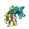
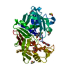
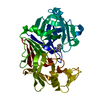
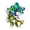
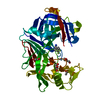
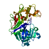
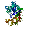
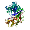
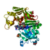
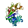
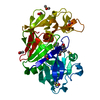
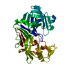
| Title | PanDDA analysis group deposition -- Endothiapepsin ground state model 18 | ||||||
 Components Components | Endothiapepsin | ||||||
 Keywords Keywords | HYDROLASE / FragMAX / FragMAXapp / fragment screening / inhibition / F2X-Entry | ||||||
| Function / homology |  Function and homology information Function and homology information | ||||||
| Biological species |  Cryphonectria parasitica (chestnut blight fungus) Cryphonectria parasitica (chestnut blight fungus) | ||||||
| Method |  X-RAY DIFFRACTION / SYNCHROTRON / FOURIER SYNTHESIS / Resolution: 1.04 Å X-RAY DIFFRACTION / SYNCHROTRON / FOURIER SYNTHESIS / Resolution: 1.04 Å | ||||||
 Authors Authors | Weiss, M.S. / Wollenhaupt, J. / Metz, A. / Barthel, T. / Lima, G.M.A. / Heine, A. / Mueller, U. / Klebe, G. | ||||||
 Citation Citation | Journal: Structure / Year: 2020 Title: F2X-Universal and F2X-Entry: Structurally Diverse Compound Libraries for Crystallographic Fragment Screening. Authors: Wollenhaupt, J. / Metz, A. / Barthel, T. / Lima, G.M.A. / Heine, A. / Mueller, U. / Klebe, G. / Weiss, M.S. | ||||||
| History |
|
- Structure visualization
| Structure viewer | Molecule: MolmilJmol/JSmol |
|---|
- Downloads & links
-Download
| PDBx/mmCIF format | 5rcw.cif.gz | 89.1 KB | Display | PDBx/mmCIF format |
|---|---|---|---|---|
| PDB format | pdb5rcw.ent.gz | 64 KB | Display | PDB format |
| PDBx/mmJSON format | 5rcw.json.gz | Tree view | PDBx/mmJSON format | |
| Others |  Other downloads Other downloads |
-Validation report
| Arichive directory | https://data.pdbj.org/pub/pdb/validation_reports/rc/5rcwftp://data.pdbj.org/pub/pdb/validation_reports/rc/5rcw | HTTPS FTP |
|---|
-Group deposition
| ID | G_1002147 (88 entries) |
|---|---|
| Title | PanDDA analysis of F2X-Entry vs. Endothiapepsin |
| Type | undefined |
| Description | PanDDA analysis of F2X-Entry vs. Endothiapepsin, including auto-refined models with ligands placed according to PanDDA-map and automatically refined models necessary to reproduce ground state model |


-Related structure data
| Similar structure data |
|---|


-Links
 PDBj
PDBj
- Assembly
| Deposited unit | 
| ||||||||
|---|---|---|---|---|---|---|---|---|---|
| 1 |
| ||||||||
| Unit cell |
|
-Components
-Protein , 1 types, 1 molecules A
| #1: Protein | Mass: 43278.664 Da / Num. of mol.: 1 / Source method: isolated from a natural source Source: (natural) Cryphonectria parasitica (chestnut blight fungus)References: UniProt: P11838, endothiapepsin |
|---|
-Non-polymers , 7 types, 343 molecules 


| #2: Chemical |  Mass: 92.094 Da / Num. of mol.: 3 / Source method: obtained synthetically / Formula: C3H8O3 Mass: 92.094 Da / Num. of mol.: 3 / Source method: obtained synthetically / Formula: C3H8O3#3: Chemical |  Mass: 59.044 Da / Num. of mol.: 2 / Source method: obtained synthetically / Formula: C2H3O2 Mass: 59.044 Da / Num. of mol.: 2 / Source method: obtained synthetically / Formula: C2H3O2#4: Chemical | ChemComp-PGE / |  Mass: 150.173 Da / Num. of mol.: 1 / Source method: obtained synthetically / Formula: C6H14O4 Mass: 150.173 Da / Num. of mol.: 1 / Source method: obtained synthetically / Formula: C6H14O4#5: Chemical | ChemComp-PG4 / |  Mass: 194.226 Da / Num. of mol.: 1 / Source method: obtained synthetically / Formula: C8H18O5 / Comment: precipitant*YM Mass: 194.226 Da / Num. of mol.: 1 / Source method: obtained synthetically / Formula: C8H18O5 / Comment: precipitant*YM#6: Chemical | ChemComp-NA / |  Mass: 22.990 Da / Num. of mol.: 1 / Source method: obtained synthetically / Formula: Na Mass: 22.990 Da / Num. of mol.: 1 / Source method: obtained synthetically / Formula: Na#7: Chemical | ChemComp-PEG / |  Mass: 106.120 Da / Num. of mol.: 1 / Source method: obtained synthetically / Formula: C4H10O3 Mass: 106.120 Da / Num. of mol.: 1 / Source method: obtained synthetically / Formula: C4H10O3#8: Water | ChemComp-HOH / | Mass: 18.015 Da / Num. of mol.: 334 / Source method: isolated from a natural source / Formula: H2O |
|---|
-Details
| Has protein modification | Y |
|---|
-Experimental details
-Experiment
| Experiment | Method: X-RAY DIFFRACTION / Number of used crystals: 1 |
|---|
- Sample preparation
| Crystal | Density Matthews: 1.89 Å3/Da / Density % sol: 35.05 % |
|---|---|
| Crystal grow | Temperature: 290 K / Method: vapor diffusion, sitting drop / pH: 4.6 Details: 0.1 M ammonium acetate, 0.1 M sodium acetate, 24-30% PEG 4000 |
-Data collection
| Diffraction | Mean temperature: 100 K | ||||||||||||||||||||||||||||||||||||||||||||||||||||||||||||||||||||||||||||||||||||||||||||||||||||||||||||||
|---|---|---|---|---|---|---|---|---|---|---|---|---|---|---|---|---|---|---|---|---|---|---|---|---|---|---|---|---|---|---|---|---|---|---|---|---|---|---|---|---|---|---|---|---|---|---|---|---|---|---|---|---|---|---|---|---|---|---|---|---|---|---|---|---|---|---|---|---|---|---|---|---|---|---|---|---|---|---|---|---|---|---|---|---|---|---|---|---|---|---|---|---|---|---|---|---|---|---|---|---|---|---|---|---|---|---|---|---|---|---|---|
| Diffraction source | Source: SYNCHROTRON / Site: MAX IV  / Beamline: BioMAX / Wavelength: 0.827 Å / Beamline: BioMAX / Wavelength: 0.827 Å | ||||||||||||||||||||||||||||||||||||||||||||||||||||||||||||||||||||||||||||||||||||||||||||||||||||||||||||||
| Detector | Type: DECTRIS EIGER X 16M / Detector: PIXEL / Date: Mar 30, 2019 | ||||||||||||||||||||||||||||||||||||||||||||||||||||||||||||||||||||||||||||||||||||||||||||||||||||||||||||||
| Radiation | Monochromator: Si(111) / Protocol: SINGLE WAVELENGTH / Monochromatic (M) / Laue (L): M / Scattering type: x-ray | ||||||||||||||||||||||||||||||||||||||||||||||||||||||||||||||||||||||||||||||||||||||||||||||||||||||||||||||
| Radiation wavelength | Wavelength: 0.827 Å / Relative weight: 1 | ||||||||||||||||||||||||||||||||||||||||||||||||||||||||||||||||||||||||||||||||||||||||||||||||||||||||||||||
| Reflection | Resolution: 1.04→42.77 Å / Num. obs: 145990 / % possible obs: 99.1 % / Redundancy: 6.818 % / Biso Wilson estimate: 15.556 Å2 / CC1/2: 0.999 / Rmerge(I) obs: 0.057 / Rrim(I) all: 0.062 / Χ2: 1.128 / Net I/σ(I): 14.15 / Num. measured all: 1048720 | ||||||||||||||||||||||||||||||||||||||||||||||||||||||||||||||||||||||||||||||||||||||||||||||||||||||||||||||
| Reflection shell | Diffraction-ID: 1
|
- Processing
| Software |
| |||||||||||||||||||||||||||||||||||||||||||||||||||||||||||||||||||||||||||
|---|---|---|---|---|---|---|---|---|---|---|---|---|---|---|---|---|---|---|---|---|---|---|---|---|---|---|---|---|---|---|---|---|---|---|---|---|---|---|---|---|---|---|---|---|---|---|---|---|---|---|---|---|---|---|---|---|---|---|---|---|---|---|---|---|---|---|---|---|---|---|---|---|---|---|---|---|
| Refinement | Method to determine structure: FOURIER SYNTHESIS / Resolution: 1.04→42.77 Å / Cor.coef. Fo:Fc: 0.98 / Cor.coef. Fo:Fc free: 0.979 / SU B: 0.415 / SU ML: 0.02 / Cross valid method: THROUGHOUT / σ(F): 0 / ESU R: 0.024 / ESU R Free: 0.024 / Stereochemistry target values: MAXIMUM LIKELIHOOD Details: HYDROGENS HAVE BEEN ADDED IN THE RIDING POSITIONS U VALUES : REFINED INDIVIDUALLY
| |||||||||||||||||||||||||||||||||||||||||||||||||||||||||||||||||||||||||||
| Solvent computation | Ion probe radii: 0.8 Å / Shrinkage radii: 0.8 Å / VDW probe radii: 1.2 Å / Solvent model: MASK | |||||||||||||||||||||||||||||||||||||||||||||||||||||||||||||||||||||||||||
| Displacement parameters | Biso max: 78.23 Å2 / Biso mean: 13.151 Å2 / Biso min: 8.02 Å2
| |||||||||||||||||||||||||||||||||||||||||||||||||||||||||||||||||||||||||||
| Refinement step | Cycle: final / Resolution: 1.04→42.77 Å
| |||||||||||||||||||||||||||||||||||||||||||||||||||||||||||||||||||||||||||
| Refine LS restraints |
| |||||||||||||||||||||||||||||||||||||||||||||||||||||||||||||||||||||||||||
| LS refinement shell | Resolution: 1.038→1.065 Å / Rfactor Rfree error: 0 / Total num. of bins used: 20
|
