 Movie
Movie Controller

[English] 日本語
 Yorodumi
- PDB-1bui: Structure of the ternary microplasmin-staphylokinase-microplasmin... -
+ Open data

- Basic information
| Entry | Database: PDB / ID: 1bui | ||||||
|---|---|---|---|---|---|---|---|
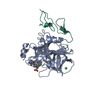
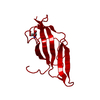
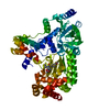
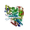
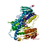
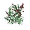
| Title | Structure of the ternary microplasmin-staphylokinase-microplasmin complex: a proteinase-cofactor-substrate complex in action | ||||||
 Components Components |
| ||||||
 Keywords Keywords | HYDROLASE/HYDROLASE INHIBITOR / PLASMIN / STAPHYLOKINASE / SERINE PROTEINASE / FIBRINOLYSIS / COFACTOR / HYDROLASE-HYDROLASE INHIBITOR complex | ||||||
| Function / homology |  Function and homology information Function and homology informationplasmin / trans-synaptic signaling by BDNF, modulating synaptic transmission / trophoblast giant cell differentiation / tissue remodeling / tissue regeneration / Signaling by PDGF / mononuclear cell migration / positive regulation of fibrinolysis / negative regulation of cell-cell adhesion mediated by cadherin / protein antigen binding ...plasmin / trans-synaptic signaling by BDNF, modulating synaptic transmission / trophoblast giant cell differentiation / tissue remodeling / tissue regeneration / Signaling by PDGF / mononuclear cell migration / positive regulation of fibrinolysis / negative regulation of cell-cell adhesion mediated by cadherin / protein antigen binding / Dissolution of Fibrin Clot / myoblast differentiation / biological process involved in interaction with symbiont / labyrinthine layer blood vessel development / muscle cell cellular homeostasis / Activation of Matrix Metalloproteinases / apolipoprotein binding / extracellular matrix disassembly / negative regulation of cell-substrate adhesion / positive regulation of blood vessel endothelial cell migration / negative regulation of fibrinolysis / fibrinolysis / Degradation of the extracellular matrix / serine-type peptidase activity / platelet alpha granule lumen / protein processing / kinase binding / Schaffer collateral - CA1 synapse / Regulation of Insulin-like Growth Factor (IGF) transport and uptake by Insulin-like Growth Factor Binding Proteins (IGFBPs) / blood coagulation / Platelet degranulation / protein-folding chaperone binding / : / protease binding / blood microparticle / endopeptidase activity / protein domain specific binding / signaling receptor binding / negative regulation of cell population proliferation / serine-type endopeptidase activity / external side of plasma membrane / glutamatergic synapse / enzyme binding / cell surface / proteolysis / extracellular space / extracellular exosome / extracellular region / plasma membrane Similarity search - Function | ||||||
| Biological species |  Homo sapiens (human) Homo sapiens (human) Staphylococcus phage 42D.m (virus) Staphylococcus phage 42D.m (virus) | ||||||
| Method |  X-RAY DIFFRACTION / SYNCHROTRON / MOLECULAR REPLACEMENT / Resolution: 2.65 Å X-RAY DIFFRACTION / SYNCHROTRON / MOLECULAR REPLACEMENT / Resolution: 2.65 Å | ||||||
 Authors Authors | Parry, M.A.A. / Fernandez-Catalan, C. / Bergner, A. / Huber, R. / Hopfner, K. / Schlott, B. / Guehrs, K. / Bode, W. | ||||||
 Citation Citation | Journal: Nat.Struct.Biol. / Year: 1998 Title: The ternary microplasmin-staphylokinase-microplasmin complex is a proteinase-cofactor-substrate complex in action. Authors: Parry, M.A. / Fernandez-Catalan, C. / Bergner, A. / Huber, R. / Hopfner, K.P. / Schlott, B. / Guhrs, K.H. / Bode, W. | ||||||
| History |
| ||||||
| Remark 650 | HELIX DETERMINATION METHOD: DSSP | ||||||
| Remark 700 | SHEET DETERMINATION METHOD: DSSP |
- Structure visualization
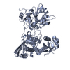
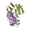
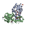
| Structure viewer | Molecule: MolmilJmol/JSmol |
|---|
- Downloads & links
-Download
| PDBx/mmCIF format | 1bui.cif.gz | 139.4 KB | Display | PDBx/mmCIF format |
|---|---|---|---|---|
| PDB format | pdb1bui.ent.gz | 107.8 KB | Display | PDB format |
| PDBx/mmJSON format | 1bui.json.gz | Tree view | PDBx/mmJSON format | |
| Others |  Other downloads Other downloads |
-Validation report
| Summary document | 1bui_validation.pdf.gz | 448.7 KB | Display | wwPDB validaton report |
|---|---|---|---|---|
| Full document | 1bui_full_validation.pdf.gz | 475.8 KB | Display | |
| Data in XML | 1bui_validation.xml.gz | 16.6 KB | Display | |
| Data in CIF | 1bui_validation.cif.gz | 26.5 KB | Display | |
| Arichive directory | https://data.pdbj.org/pub/pdb/validation_reports/bu/1buiftp://data.pdbj.org/pub/pdb/validation_reports/bu/1bui | HTTPS FTP |
-Related structure data


-Links
 PDBj
PDBj


- Assembly
| Deposited unit | 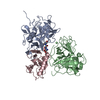
| ||||||||
|---|---|---|---|---|---|---|---|---|---|
| 1 |
| ||||||||
| Unit cell |
|
-Components
| #1: Protein | Mass: 27335.402 Da / Num. of mol.: 2 / Fragment: Peptidase S1 catalytic domain Source method: isolated from a genetically manipulated source Source: (gene. exp.) Homo sapiens (human) / Gene: PLG / Production host:  #2: Protein | | Mass: 14598.640 Da / Num. of mol.: 1 / Source method: isolated from a natural source / Source: (natural) Staphylococcus phage 42D.m (virus) / Genus: Lambda-like viruses / References: UniProt: P15240#3: Chemical | ChemComp-0GJ / | 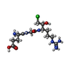  Type: peptide-like, Peptide-like / Class: Inhibitor / Mass: 395.862 Da / Num. of mol.: 1 / Source method: obtained synthetically / Formula: C14H28ClN6O5 / References: GLU-GLY-ARG-CHLOROMETHYL KETONE Type: peptide-like, Peptide-like / Class: Inhibitor / Mass: 395.862 Da / Num. of mol.: 1 / Source method: obtained synthetically / Formula: C14H28ClN6O5 / References: GLU-GLY-ARG-CHLOROMETHYL KETONE#4: Water | ChemComp-HOH / |  Mass: 18.015 Da / Num. of mol.: 313 / Source method: isolated from a natural source / Formula: H2O Mass: 18.015 Da / Num. of mol.: 313 / Source method: isolated from a natural source / Formula: H2OHas protein modification | Y | Nonpolymer details | THE UNBOUND FORM OF THE INHIBITOR IS GLU-GLY-ARG-CMK-CHLOROMETHYLKETONE. UPON REACTION WITH PROTEIN ...THE UNBOUND FORM OF THE INHIBITOR IS GLU-GLY-ARG-CMK-CHLOROMETH | |
|---|
-Experimental details
-Experiment
| Experiment | Method: X-RAY DIFFRACTION / Number of used crystals: 1 |
|---|
- Sample preparation
| Crystal | Density Matthews: 3.42 Å3/Da / Density % sol: 67 % | |||||||||||||||
|---|---|---|---|---|---|---|---|---|---|---|---|---|---|---|---|---|
| Crystal grow | pH: 7.5 / Details: pH 7.5 | |||||||||||||||
| Crystal | *PLUS | |||||||||||||||
| Crystal grow | *PLUS Method: vapor diffusion, sitting drop | |||||||||||||||
| Components of the solutions | *PLUS
|
-Data collection
| Diffraction | Mean temperature: 190 K |
|---|---|
| Diffraction source | Source: SYNCHROTRON / Site: MPG/DESY, HAMBURG  / Beamline: BW6 / Wavelength: 1.06 / Beamline: BW6 / Wavelength: 1.06 |
| Detector | Type: MARRESEARCH / Detector: CCD / Date: May 15, 1998 |
| Radiation | Protocol: SINGLE WAVELENGTH / Monochromatic (M) / Laue (L): M / Scattering type: x-ray |
| Radiation wavelength | Wavelength: 1.06 Å / Relative weight: 1 |
| Reflection | Resolution: 2.65→10 Å / Num. obs: 28745 / % possible obs: 99.5 % / Observed criterion σ(I): 0 / Redundancy: 2 % / Rmerge(I) obs: 0.075 |
| Reflection | *PLUS Num. measured all: 483198 |
| Reflection shell | *PLUS % possible obs: 98 % |
- Processing
| Software |
| ||||||||||||||||||||||||||||||||||||||||||||||||||||||||||||
|---|---|---|---|---|---|---|---|---|---|---|---|---|---|---|---|---|---|---|---|---|---|---|---|---|---|---|---|---|---|---|---|---|---|---|---|---|---|---|---|---|---|---|---|---|---|---|---|---|---|---|---|---|---|---|---|---|---|---|---|---|---|
| Refinement | Method to determine structure: MOLECULAR REPLACEMENT Starting model: PDB ENTRIES 1FAX AND 2SAK Resolution: 2.65→10 Å / σ(F): 0
| ||||||||||||||||||||||||||||||||||||||||||||||||||||||||||||
| Refinement step | Cycle: LAST / Resolution: 2.65→10 Å
| ||||||||||||||||||||||||||||||||||||||||||||||||||||||||||||
| Refine LS restraints |
| ||||||||||||||||||||||||||||||||||||||||||||||||||||||||||||
| Software | *PLUS Name: X-PLOR / Classification: refinement | ||||||||||||||||||||||||||||||||||||||||||||||||||||||||||||
| Refinement | *PLUS Lowest resolution: 10 Å / σ(F): 0 / % reflection Rfree: 7 % | ||||||||||||||||||||||||||||||||||||||||||||||||||||||||||||
| Solvent computation | *PLUS | ||||||||||||||||||||||||||||||||||||||||||||||||||||||||||||
| Displacement parameters | *PLUS |
