 Movie
Movie Controller

[English] 日本語
 Yorodumi
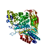
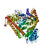
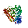
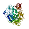
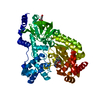
- PDB-2x42: Structure of beta-glucosidase 3B from Thermotoga neapolitana in c... -
+ Open data

- Basic information
| Entry | Database: PDB / ID: 2x42 | ||||||
|---|---|---|---|---|---|---|---|
| Title | Structure of beta-glucosidase 3B from Thermotoga neapolitana in complex with alpha-D-glucose | ||||||
 Components Components | BETA-GLUCOSIDASE | ||||||
 Keywords Keywords | HYDROLASE / TIM BARREL FOLD / FIBRONECTIN TYPE III FOLD | ||||||
| Function / homology |  Function and homology information Function and homology informationbeta-glucosidase / beta-glucosidase activity / carbohydrate metabolic process Similarity search - Function | ||||||
| Biological species |   THERMOTOGA NEAPOLITANA (bacteria) THERMOTOGA NEAPOLITANA (bacteria) | ||||||
| Method |  X-RAY DIFFRACTION / SYNCHROTRON / MOLECULAR REPLACEMENT / Resolution: 2.099 Å X-RAY DIFFRACTION / SYNCHROTRON / MOLECULAR REPLACEMENT / Resolution: 2.099 Å | ||||||
 Authors Authors | Pozzo, T. / Karlsson, E.N. / Logan, D.T. | ||||||
 Citation Citation | Journal: J.Mol.Biol. / Year: 2010 Title: Structural and Functional Analysis of Beta-Glucosidase 3B from Thermotoga Neapolitana: A Thermostable 3-Domain Representative of Glycoside Hydrolase Family 3 Authors: Pozzo, T. / Linares Pasten, J. / Karlsson, E.N. / Logan, D.T. #1: Journal: Acta Crystallogr.,Sect.F / Year: 2007 Title: Expression, Purification, Crystallization and Preliminary X-Ray Diffraction Analysis of Thermotoga Neapolitana Beta-Glucosidase B. Authors: Turner, P. / Pramhed, A. / Kanders, E. / Hedstrom, M. / Karlsson, E.N. / Logan, D.T. | ||||||
| History |
|
- Structure visualization
| Structure viewer | Molecule: MolmilJmol/JSmol |
|---|
- Downloads & links
-Download
| PDBx/mmCIF format | 2x42.cif.gz | 299.7 KB | Display | PDBx/mmCIF format |
|---|---|---|---|---|
| PDB format | pdb2x42.ent.gz | 242.8 KB | Display | PDB format |
| PDBx/mmJSON format | 2x42.json.gz | Tree view | PDBx/mmJSON format | |
| Others |  Other downloads Other downloads |
-Validation report
| Arichive directory | https://data.pdbj.org/pub/pdb/validation_reports/x4/2x42ftp://data.pdbj.org/pub/pdb/validation_reports/x4/2x42 | HTTPS FTP |
|---|
-Related structure data
| Related structure data |  2x40SC 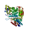 2x41C  2wt3 2wt5 2wt6 S: Starting model for refinement C: citing same article ( |
|---|---|
| Similar structure data |


-Links
 PDBj
PDBj


- Assembly
| Deposited unit | 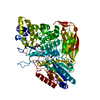
| ||||||||
|---|---|---|---|---|---|---|---|---|---|
| 1 |
| ||||||||
| Unit cell |
|
-Components
| #1: Protein | Mass: 81217.852 Da / Num. of mol.: 1 / Mutation: YES Source method: isolated from a genetically manipulated source Source: (gene. exp.) THERMOTOGA NEAPOLITANA (bacteria) / Strain: DSM 4359 / Description: GERMAN COLLECTION OF MICROORGANISMS (DSM) / Production host: | ||||
|---|---|---|---|---|---|
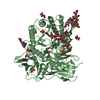
| #2: Sugar | ChemComp-GLC /   Type: D-saccharide, alpha linking / Mass: 180.156 Da / Num. of mol.: 1 Type: D-saccharide, alpha linking / Mass: 180.156 Da / Num. of mol.: 1Source method: isolated from a genetically manipulated source Formula: C6H12O6 | ||||
| #3: Chemical | ChemComp-BR /   Mass: 79.904 Da / Num. of mol.: 4 / Source method: obtained synthetically / Formula: Br Mass: 79.904 Da / Num. of mol.: 4 / Source method: obtained synthetically / Formula: Br#4: Water | ChemComp-HOH / |  Mass: 18.015 Da / Num. of mol.: 442 / Source method: isolated from a natural source / Formula: H2O Mass: 18.015 Da / Num. of mol.: 442 / Source method: isolated from a natural source / Formula: H2OCompound details | ENGINEERED | |
-Experimental details
-Experiment
| Experiment | Method: X-RAY DIFFRACTION / Number of used crystals: 1 |
|---|
- Sample preparation
| Crystal | Density Matthews: 2.57 Å3/Da / Density % sol: 57 % / Description: NONE |
|---|---|
| Crystal grow | Temperature: 293 K / pH: 7.4 Details: 3-5 MG/ML PROTEIN, 16-18 % (V/V) PEG 3350, 0.2 M NABR, 90 MM BIS-TRIS PROPANE PH 7.4, 20C |
-Data collection
| Diffraction | Mean temperature: 100 K |
|---|---|
| Diffraction source | Source: SYNCHROTRON / Site: MAX II  / Beamline: I911-2 / Wavelength: 1.0379 / Beamline: I911-2 / Wavelength: 1.0379 |
| Detector | Type: MARRESEARCH SX-165 / Detector: CCD / Date: Sep 24, 2008 / Details: MULTILAYER MIRRORS |
| Radiation | Monochromator: SI(311) / Protocol: SINGLE WAVELENGTH / Monochromatic (M) / Laue (L): M / Scattering type: x-ray |
| Radiation wavelength | Wavelength: 1.0379 Å / Relative weight: 1 |
| Reflection | Resolution: 2.1→29.2 Å / Num. obs: 47706 / % possible obs: 96.5 % / Observed criterion σ(I): 0 / Redundancy: 4.6 % / Biso Wilson estimate: 32.69 Å2 / Rmerge(I) obs: 0.07 / Net I/σ(I): 15.4 |
| Reflection shell | Resolution: 2.1→2.2 Å / Rmerge(I) obs: 0.56 / Mean I/σ(I) obs: 2.2 / % possible all: 78.3 |
- Processing
| Software |
| ||||||||||||||||||||||||||||||||||||||||||||||||||||||||||||||||||||||||||||||||||||||||||||||||||||||||||||||||||||||||||||||
|---|---|---|---|---|---|---|---|---|---|---|---|---|---|---|---|---|---|---|---|---|---|---|---|---|---|---|---|---|---|---|---|---|---|---|---|---|---|---|---|---|---|---|---|---|---|---|---|---|---|---|---|---|---|---|---|---|---|---|---|---|---|---|---|---|---|---|---|---|---|---|---|---|---|---|---|---|---|---|---|---|---|---|---|---|---|---|---|---|---|---|---|---|---|---|---|---|---|---|---|---|---|---|---|---|---|---|---|---|---|---|---|---|---|---|---|---|---|---|---|---|---|---|---|---|---|---|---|
| Refinement | Method to determine structure: MOLECULAR REPLACEMENT Starting model: PDB ENTRY 2X40 Resolution: 2.099→29.275 Å / SU ML: 0.31 / σ(F): 1.35 / Phase error: 21.65 / Stereochemistry target values: ML
| ||||||||||||||||||||||||||||||||||||||||||||||||||||||||||||||||||||||||||||||||||||||||||||||||||||||||||||||||||||||||||||||
| Solvent computation | Shrinkage radii: 0.9 Å / VDW probe radii: 1.11 Å / Solvent model: FLAT BULK SOLVENT MODEL / Bsol: 44.433 Å2 / ksol: 0.353 e/Å3 | ||||||||||||||||||||||||||||||||||||||||||||||||||||||||||||||||||||||||||||||||||||||||||||||||||||||||||||||||||||||||||||||
| Displacement parameters | Biso mean: 36.8 Å2
| ||||||||||||||||||||||||||||||||||||||||||||||||||||||||||||||||||||||||||||||||||||||||||||||||||||||||||||||||||||||||||||||
| Refinement step | Cycle: LAST / Resolution: 2.099→29.275 Å
| ||||||||||||||||||||||||||||||||||||||||||||||||||||||||||||||||||||||||||||||||||||||||||||||||||||||||||||||||||||||||||||||
| Refine LS restraints |
| ||||||||||||||||||||||||||||||||||||||||||||||||||||||||||||||||||||||||||||||||||||||||||||||||||||||||||||||||||||||||||||||
| LS refinement shell |
| ||||||||||||||||||||||||||||||||||||||||||||||||||||||||||||||||||||||||||||||||||||||||||||||||||||||||||||||||||||||||||||||
| Refinement TLS params. | Method: refined / Refine-ID: X-RAY DIFFRACTION
| ||||||||||||||||||||||||||||||||||||||||||||||||||||||||||||||||||||||||||||||||||||||||||||||||||||||||||||||||||||||||||||||
| Refinement TLS group |
|
