 Movie
Movie Controller


+ Open data

- Basic information
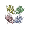
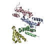
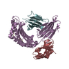
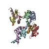
| Entry | Database: PDB / ID: 1bu1 | ||||||
|---|---|---|---|---|---|---|---|
| Title | SRC FAMILY KINASE HCK SH3 DOMAIN | ||||||
 Components Components | PROTEIN (HEMOPOIETIC CELL KINASE) | ||||||
 Keywords Keywords | TRANSFERASE / TYROSINE-PROTEIN KINASE / SIGNAL TRANSDUCTION / SH3 | ||||||
| Function / homology |  Function and homology information Function and homology informationleukocyte degranulation / leukocyte migration involved in immune response / respiratory burst after phagocytosis / regulation of podosome assembly / innate immune response-activating signaling pathway / : / FLT3 signaling through SRC family kinases / regulation of phagocytosis / Nef and signal transduction / Fc-gamma receptor signaling pathway involved in phagocytosis ...leukocyte degranulation / leukocyte migration involved in immune response / respiratory burst after phagocytosis / regulation of podosome assembly / innate immune response-activating signaling pathway / : / FLT3 signaling through SRC family kinases / regulation of phagocytosis / Nef and signal transduction / Fc-gamma receptor signaling pathway involved in phagocytosis / mesoderm development / positive regulation of actin filament polymerization / FCGR activation / type II interferon-mediated signaling pathway / transport vesicle / Signaling by CSF3 (G-CSF) / cell projection / phosphotyrosine residue binding / peptidyl-tyrosine phosphorylation / lipopolysaccharide-mediated signaling pathway / FCGR3A-mediated IL10 synthesis / cell surface receptor protein tyrosine kinase signaling pathway / integrin-mediated signaling pathway / regulation of actin cytoskeleton organization / non-specific protein-tyrosine kinase / FCGR3A-mediated phagocytosis / Regulation of signaling by CBL / negative regulation of inflammatory response to antigenic stimulus / non-membrane spanning protein tyrosine kinase activity / caveola / Inactivation of CSF3 (G-CSF) signaling / cytoplasmic side of plasma membrane / cytokine-mediated signaling pathway / Signaling by CSF1 (M-CSF) in myeloid cells / protein autophosphorylation / regulation of cell shape / protein tyrosine kinase activity / regulation of inflammatory response / cytoskeleton / protein phosphorylation / cell differentiation / lysosome / cell adhesion / intracellular signal transduction / defense response to Gram-positive bacterium / inflammatory response / signaling receptor binding / focal adhesion / positive regulation of cell population proliferation / lipid binding / negative regulation of apoptotic process / Golgi apparatus / ATP binding / nucleus / plasma membrane / cytosol Similarity search - Function | ||||||
| Biological species |  Homo sapiens (human) Homo sapiens (human) | ||||||
| Method |  X-RAY DIFFRACTION / SYNCHROTRON / MOLECULAR REPLACEMENT / Resolution: 2.6 Å X-RAY DIFFRACTION / SYNCHROTRON / MOLECULAR REPLACEMENT / Resolution: 2.6 Å | ||||||
 Authors Authors | Arold, S. / Franken, P. / Dumas, C. | ||||||
 Citation Citation | Journal: Biochemistry / Year: 1998 Title: RT loop flexibility enhances the specificity of Src family SH3 domains for HIV-1 Nef. Authors: Arold, S. / O'Brien, R. / Franken, P. / Strub, M.P. / Hoh, F. / Dumas, C. / Ladbury, J.E. | ||||||
| History |
|
- Structure visualization
| Structure viewer | Molecule: MolmilJmol/JSmol |
|---|
- Downloads & links
-Download
| PDBx/mmCIF format | 1bu1.cif.gz | 80.1 KB | Display | PDBx/mmCIF format |
|---|---|---|---|---|
| PDB format | pdb1bu1.ent.gz | 61.6 KB | Display | PDB format |
| PDBx/mmJSON format | 1bu1.json.gz | Tree view | PDBx/mmJSON format | |
| Others |  Other downloads Other downloads |
-Validation report
| Arichive directory | https://data.pdbj.org/pub/pdb/validation_reports/bu/1bu1ftp://data.pdbj.org/pub/pdb/validation_reports/bu/1bu1 | HTTPS FTP |
|---|
-Related structure data
| Related structure data |  2hckS S: Starting model for refinement |
|---|---|
| Similar structure data |


-Links
 PDBj
PDBj


- Assembly
| Deposited unit | 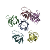
| ||||||||||||||||||||||||||||
|---|---|---|---|---|---|---|---|---|---|---|---|---|---|---|---|---|---|---|---|---|---|---|---|---|---|---|---|---|---|
| 1 |
| ||||||||||||||||||||||||||||
| Unit cell |
| ||||||||||||||||||||||||||||
| Noncrystallographic symmetry (NCS) | NCS oper:
|
-Components
| #1: Protein | Mass: 6635.428 Da / Num. of mol.: 6 / Fragment: SH3 Source method: isolated from a genetically manipulated source Source: (gene. exp.) Homo sapiens (human) / Cell line: BL21 (DE3) / Gene: HUMAN HCK / Species (production host): Escherichia coli / Gene (production host): HUMAN HCK / Production host:  #2: Water | ChemComp-HOH / |  Mass: 18.015 Da / Num. of mol.: 70 / Source method: isolated from a natural source / Formula: H2O Mass: 18.015 Da / Num. of mol.: 70 / Source method: isolated from a natural source / Formula: H2O |
|---|
-Experimental details
-Experiment
| Experiment | Method: X-RAY DIFFRACTION / Number of used crystals: 2 |
|---|
- Sample preparation
| Crystal | Density Matthews: 2.7 Å3/Da / Density % sol: 52.7 % | ||||||||||||||||||||||||||||||||||||||||||||||||
|---|---|---|---|---|---|---|---|---|---|---|---|---|---|---|---|---|---|---|---|---|---|---|---|---|---|---|---|---|---|---|---|---|---|---|---|---|---|---|---|---|---|---|---|---|---|---|---|---|---|
| Crystal grow | Temperature: 294 K / Method: vapor diffusion, hanging drop / pH: 9.3 Details: HANGING DROPS (2UL) OF 4.3MG/ML PROTEIN WERE MIXED WITH EQUAL VOLUMES OF RESERVOIR BUFFER CONTAINING 3.7 M SODIUM FORMATE, 2% PEG 3000, 100 MM BICINE (PH 9.3). THE MIXED DROPS WERE STORED AT ...Details: HANGING DROPS (2UL) OF 4.3MG/ML PROTEIN WERE MIXED WITH EQUAL VOLUMES OF RESERVOIR BUFFER CONTAINING 3.7 M SODIUM FORMATE, 2% PEG 3000, 100 MM BICINE (PH 9.3). THE MIXED DROPS WERE STORED AT 21 DEGREES, vapor diffusion - hanging drop, temperature 294K | ||||||||||||||||||||||||||||||||||||||||||||||||
| Crystal | *PLUS | ||||||||||||||||||||||||||||||||||||||||||||||||
| Crystal grow | *PLUS Temperature: 21 ℃ / pH: 8 / Method: vapor diffusion, hanging drop | ||||||||||||||||||||||||||||||||||||||||||||||||
| Components of the solutions | *PLUS
|
-Data collection
| Diffraction | Mean temperature: 280 K |
|---|---|
| Diffraction source | Source: SYNCHROTRON / Site: ESRF  / Beamline: BM02 / Wavelength: 1.074 / Beamline: BM02 / Wavelength: 1.074 |
| Detector | Type: PRINCETON / THOMSON / Detector: CCD / Date: Nov 15, 1996 / Details: TWO BENT MIRRORS |
| Radiation | Monochromator: TWO SILICON CRYSTALS / Protocol: SINGLE WAVELENGTH / Monochromatic (M) / Laue (L): M / Scattering type: x-ray |
| Radiation wavelength | Wavelength: 1.074 Å / Relative weight: 1 |
| Reflection | Resolution: 2.6→34 Å / Num. obs: 13043 / % possible obs: 93 % / Observed criterion σ(I): 0 / Redundancy: 4.8 % / Rmerge(I) obs: 0.053 / Rsym value: 0.053 / Net I/σ(I): 35 |
| Reflection shell | Resolution: 2.6→2.7 Å / Redundancy: 10.2 % / Rmerge(I) obs: 0.189 / Rsym value: 0.189 / % possible all: 62.1 |
| Reflection shell | *PLUS Lowest resolution: 2.72 Å / % possible obs: 62 % |
- Processing
| Software |
| ||||||||||||||||||||||||||||||||||||||||||||||||||||||||||||
|---|---|---|---|---|---|---|---|---|---|---|---|---|---|---|---|---|---|---|---|---|---|---|---|---|---|---|---|---|---|---|---|---|---|---|---|---|---|---|---|---|---|---|---|---|---|---|---|---|---|---|---|---|---|---|---|---|---|---|---|---|---|
| Refinement | Method to determine structure: MOLECULAR REPLACEMENT Starting model: 2HCK Resolution: 2.6→34 Å / Data cutoff high absF: 10000000 / Data cutoff low absF: 0.001 / Cross valid method: THROUGHOUT / σ(F): 0
| ||||||||||||||||||||||||||||||||||||||||||||||||||||||||||||
| Displacement parameters | Biso mean: 30.4 Å2
| ||||||||||||||||||||||||||||||||||||||||||||||||||||||||||||
| Refinement step | Cycle: LAST / Resolution: 2.6→34 Å
| ||||||||||||||||||||||||||||||||||||||||||||||||||||||||||||
| Refine LS restraints |
| ||||||||||||||||||||||||||||||||||||||||||||||||||||||||||||
| Refine LS restraints NCS | NCS model details: RESTRAINED | ||||||||||||||||||||||||||||||||||||||||||||||||||||||||||||
| LS refinement shell | Resolution: 2.6→2.67 Å / Total num. of bins used: 13 | ||||||||||||||||||||||||||||||||||||||||||||||||||||||||||||
| Xplor file | Serial no: 1 / Param file: PARAM19X.PRO / Topol file: TOPH19X.PRO | ||||||||||||||||||||||||||||||||||||||||||||||||||||||||||||
| Software | *PLUS Name: X-PLOR / Version: 3.8 / Classification: refinement | ||||||||||||||||||||||||||||||||||||||||||||||||||||||||||||
| Refinement | *PLUS Highest resolution: 2.6 Å / Lowest resolution: 34 Å / σ(F): 0 / % reflection Rfree: 4.5 % | ||||||||||||||||||||||||||||||||||||||||||||||||||||||||||||
| Solvent computation | *PLUS | ||||||||||||||||||||||||||||||||||||||||||||||||||||||||||||
| Displacement parameters | *PLUS Biso mean: 30.4 Å2 | ||||||||||||||||||||||||||||||||||||||||||||||||||||||||||||
| Refine LS restraints | *PLUS
| ||||||||||||||||||||||||||||||||||||||||||||||||||||||||||||
| LS refinement shell | *PLUS Highest resolution: 2.6 Å |
