 Movie
Movie Controller


+ Open data

- Basic information
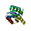
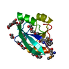
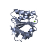
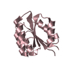
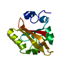
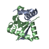
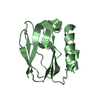
| Entry | Database: PDB / ID: 1bhd | ||||||
|---|---|---|---|---|---|---|---|
| Title | SECOND CALPONIN HOMOLOGY DOMAIN FROM UTROPHIN | ||||||
 Components Components | UTROPHIN | ||||||
 Keywords Keywords | STRUCTURAL PROTEIN / CALPONIN HOMOLOGY / ACTIN BINDING | ||||||
| Function / homology |  Function and homology information Function and homology informationsynaptic signaling / dystrophin-associated glycoprotein complex / vinculin binding / EGR2 and SOX10-mediated initiation of Schwann cell myelination / regulation of sodium ion transmembrane transport / Formation of the dystrophin-glycoprotein complex (DGC) / filopodium membrane / positive regulation of cell-matrix adhesion / muscle organ development / muscle contraction ...synaptic signaling / dystrophin-associated glycoprotein complex / vinculin binding / EGR2 and SOX10-mediated initiation of Schwann cell myelination / regulation of sodium ion transmembrane transport / Formation of the dystrophin-glycoprotein complex (DGC) / filopodium membrane / positive regulation of cell-matrix adhesion / muscle organ development / muscle contraction / filopodium / neuromuscular junction / sarcolemma / integrin binding / actin binding / postsynaptic membrane / cytoskeleton / cilium / ciliary basal body / protein kinase binding / protein-containing complex / extracellular exosome / zinc ion binding / nucleoplasm / membrane / plasma membrane / cytosol / cytoplasm Similarity search - Function | ||||||
| Biological species |  Homo sapiens (human) Homo sapiens (human) | ||||||
| Method |  X-RAY DIFFRACTION / SYNCHROTRON / MOLECULAR REPLACEMENT / Resolution: 2 Å X-RAY DIFFRACTION / SYNCHROTRON / MOLECULAR REPLACEMENT / Resolution: 2 Å | ||||||
 Authors Authors | Keep, N.H. / Winder, S.J. / Kendrick-Jones, J. | ||||||
 Citation Citation | Journal: J.Mol.Biol. / Year: 1999 Title: The 2.0 A structure of the second calponin homology domain from the actin-binding region of the dystrophin homologue utrophin. Authors: Keep, N.H. / Norwood, F.L. / Moores, C.A. / Winder, S.J. / Kendrick-Jones, J. | ||||||
| History |
|
- Structure visualization
| Structure viewer | Molecule: MolmilJmol/JSmol |
|---|
- Downloads & links
-Download
| PDBx/mmCIF format | 1bhd.cif.gz | 62 KB | Display | PDBx/mmCIF format |
|---|---|---|---|---|
| PDB format | pdb1bhd.ent.gz | 44 KB | Display | PDB format |
| PDBx/mmJSON format | 1bhd.json.gz | Tree view | PDBx/mmJSON format | |
| Others |  Other downloads Other downloads |
-Validation report
| Summary document | 1bhd_validation.pdf.gz | 436.7 KB | Display | wwPDB validaton report |
|---|---|---|---|---|
| Full document | 1bhd_full_validation.pdf.gz | 442.1 KB | Display | |
| Data in XML | 1bhd_validation.xml.gz | 12.8 KB | Display | |
| Data in CIF | 1bhd_validation.cif.gz | 17.8 KB | Display | |
| Arichive directory | https://data.pdbj.org/pub/pdb/validation_reports/bh/1bhdftp://data.pdbj.org/pub/pdb/validation_reports/bh/1bhd | HTTPS FTP |
-Related structure data
| Related structure data | 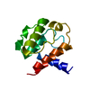 1aa2S S: Starting model for refinement |
|---|---|
| Similar structure data |


-Links
 PDBj
PDBj


- Assembly
| Deposited unit | 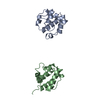
| ||||||||
|---|---|---|---|---|---|---|---|---|---|
| 1 | 
| ||||||||
| 2 | 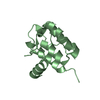
| ||||||||
| Unit cell |
| ||||||||
| Noncrystallographic symmetry (NCS) | NCS oper: (Code: given Matrix: (-0.601644, -0.050091, -0.797192), Vector: |
-Components
| #1: Protein | Mass: 13678.681 Da / Num. of mol.: 2 Fragment: 2ND CALPONIN HOMOLOGY DOMAIN FROM ACTIN BINDING REGION Source method: isolated from a genetically manipulated source Source: (gene. exp.) Homo sapiens (human) / Tissue: MUSCLE / Description: EXPRESSED IN ESCHERICHIA COLI / Cell line: BL21 / Cellular location: LINKS CYTOSKELETON TO PLASMA MEMBRANE / Gene: UTRN, DMDL / Organ: PLASMA / Plasmid: PSJW1 (T7) / Species (production host): Escherichia coli / Cellular location (production host): CYTOPLASM / Gene (production host): UTRN DMDL / Production host:  #2: Water | ChemComp-HOH / |  Mass: 18.015 Da / Num. of mol.: 173 / Source method: isolated from a natural source / Formula: H2O Mass: 18.015 Da / Num. of mol.: 173 / Source method: isolated from a natural source / Formula: H2O |
|---|
-Experimental details
-Experiment
| Experiment | Method: X-RAY DIFFRACTION / Number of used crystals: 1 |
|---|
- Sample preparation
| Crystal | Density Matthews: 2.2 Å3/Da / Density % sol: 44.24 % | |||||||||||||||||||||||||
|---|---|---|---|---|---|---|---|---|---|---|---|---|---|---|---|---|---|---|---|---|---|---|---|---|---|---|
| Crystal grow | pH: 7.6 / Details: pH 7.6 | |||||||||||||||||||||||||
| Crystal grow | *PLUS Temperature: 20 ℃ / Method: vapor diffusion | |||||||||||||||||||||||||
| Components of the solutions | *PLUS
|
-Data collection
| Diffraction | Mean temperature: 100 K |
|---|---|
| Diffraction source | Source: SYNCHROTRON / Site: SRS  / Beamline: PX9.5 / Wavelength: 1 / Beamline: PX9.5 / Wavelength: 1 |
| Detector | Type: MARRESEARCH / Detector: IMAGE PLATE / Date: Aug 25, 1997 |
| Radiation | Monochromator: SI(111) / Monochromatic (M) / Laue (L): M / Scattering type: x-ray |
| Radiation wavelength | Wavelength: 1 Å / Relative weight: 1 |
| Reflection | Resolution: 2→22 Å / Num. obs: 14514 / % possible obs: 99.5 % / Observed criterion σ(I): 0 / Redundancy: 2.8 % / Biso Wilson estimate: 23.85 Å2 / Rmerge(I) obs: 0.032 / Net I/σ(I): 10.8 |
| Reflection shell | Resolution: 1.99→2.1 Å / Redundancy: 2.8 % / Rmerge(I) obs: 0.125 / Mean I/σ(I) obs: 5.9 / % possible all: 98.9 |
| Reflection | *PLUS Num. obs: 16459 / Num. measured all: 46728 |
| Reflection shell | *PLUS % possible obs: 98.9 % / Num. unique obs: 2296 / Num. measured obs: 6400 |
- Processing
| Software |
| ||||||||||||||||||||||||||||||||||||||||||||||||||||||||||||||||||||||||||||||||||||
|---|---|---|---|---|---|---|---|---|---|---|---|---|---|---|---|---|---|---|---|---|---|---|---|---|---|---|---|---|---|---|---|---|---|---|---|---|---|---|---|---|---|---|---|---|---|---|---|---|---|---|---|---|---|---|---|---|---|---|---|---|---|---|---|---|---|---|---|---|---|---|---|---|---|---|---|---|---|---|---|---|---|---|---|---|---|
| Refinement | Method to determine structure: MOLECULAR REPLACEMENT Starting model: PDB 1AA2 Resolution: 2→20 Å / Cross valid method: THROUGHOUT / σ(F): 0 Details: 0.011 AROMATIC PLANAR DEVIATION VALUE ABOVE IS FOR PEPTIDE. ESTIMATED COORDINATE ERROR. ESD FROM SIGMAA (A) : 0.185 LOW RESOLUTION CUTOFF (A) : 20.0
| ||||||||||||||||||||||||||||||||||||||||||||||||||||||||||||||||||||||||||||||||||||
| Displacement parameters | Biso mean: 31.23 Å2
| ||||||||||||||||||||||||||||||||||||||||||||||||||||||||||||||||||||||||||||||||||||
| Refine analyze | Luzzati d res low obs: 20 Å / Luzzati sigma a obs: 0.18 Å | ||||||||||||||||||||||||||||||||||||||||||||||||||||||||||||||||||||||||||||||||||||
| Refinement step | Cycle: LAST / Resolution: 2→20 Å
| ||||||||||||||||||||||||||||||||||||||||||||||||||||||||||||||||||||||||||||||||||||
| Refine LS restraints |
| ||||||||||||||||||||||||||||||||||||||||||||||||||||||||||||||||||||||||||||||||||||
| Software | *PLUS Name: REFMAC / Classification: refinement | ||||||||||||||||||||||||||||||||||||||||||||||||||||||||||||||||||||||||||||||||||||
| Refinement | *PLUS Rfactor obs: 0.185 | ||||||||||||||||||||||||||||||||||||||||||||||||||||||||||||||||||||||||||||||||||||
| Solvent computation | *PLUS | ||||||||||||||||||||||||||||||||||||||||||||||||||||||||||||||||||||||||||||||||||||
| Displacement parameters | *PLUS | ||||||||||||||||||||||||||||||||||||||||||||||||||||||||||||||||||||||||||||||||||||
| LS refinement shell | *PLUS Highest resolution: 2 Å / Lowest resolution: 2.13 Å / Rfactor Rfree: 0.319 / Num. reflection Rfree: 132 / % reflection Rfree: 5 % / Rfactor obs: 0.188 |
