 Movie
Movie Controller

[English] 日本語
 Yorodumi
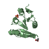
- PDB-6l8g: High resolution structure of YoeB in complex with YefM C-terminus... -
+ Open data

- Basic information
| Entry | Database: PDB / ID: 6l8g | ||||||
|---|---|---|---|---|---|---|---|
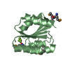
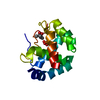
| Title | High resolution structure of YoeB in complex with YefM C-terminus(46N-83V) from Staphylococcus aureus. | ||||||
 Components Components |
| ||||||
 Keywords Keywords | ANTITOXIN/TOXIN / toxin-antitoxin / microbial RNase / YoeB / TOXIN / ANTITOXIN-TOXIN complex | ||||||
| Function / homology |  Function and homology information Function and homology informationRNA catabolic process / endonuclease activity / sequence-specific DNA binding / DNA-binding transcription factor activity / negative regulation of DNA-templated transcription / hydrolase activity / regulation of DNA-templated transcription / RNA binding Similarity search - Function | ||||||
| Biological species |   Staphylococcus aureus (bacteria) Staphylococcus aureus (bacteria) | ||||||
| Method |  X-RAY DIFFRACTION / SYNCHROTRON / MOLECULAR REPLACEMENT / Resolution: 1 Å X-RAY DIFFRACTION / SYNCHROTRON / MOLECULAR REPLACEMENT / Resolution: 1 Å | ||||||
 Authors Authors | Yue, J. / Xue, L. | ||||||
| Funding support |  China, 1items China, 1items
| ||||||
 Citation Citation | Journal: To Be Published Title: Structural insight into the mechanism of conditional cooperativity in the YoeB-YefM toxin-antitoxin system Authors: Xue, L. / Yue, J. | ||||||
| History |
|
- Structure visualization
| Structure viewer | Molecule: MolmilJmol/JSmol |
|---|
- Downloads & links
-Download
| PDBx/mmCIF format | 6l8g.cif.gz | 50.3 KB | Display | PDBx/mmCIF format |
|---|---|---|---|---|
| PDB format | pdb6l8g.ent.gz | 28.5 KB | Display | PDB format |
| PDBx/mmJSON format | 6l8g.json.gz | Tree view | PDBx/mmJSON format | |
| Others |  Other downloads Other downloads |
-Validation report
| Arichive directory | https://data.pdbj.org/pub/pdb/validation_reports/l8/6l8gftp://data.pdbj.org/pub/pdb/validation_reports/l8/6l8g | HTTPS FTP |
|---|
-Related structure data
| Similar structure data |
|---|


-Links
 PDBj
PDBj- Assembly
| Deposited unit | 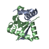
| ||||||||||||
|---|---|---|---|---|---|---|---|---|---|---|---|---|---|
| 1 |
| ||||||||||||
| Unit cell |
|
-Components
| #1: Protein/peptide | Mass: 4306.786 Da / Num. of mol.: 1 / Fragment: C-terminus Source method: isolated from a genetically manipulated source Source: (gene. exp.) Staphylococcus aureus (strain NCTC 8325) (bacteria)Strain: NCTC 8325 / Gene: SAOUHSC_02692 / Production host: |
|---|---|
| #2: Protein | Mass: 10457.985 Da / Num. of mol.: 1 Source method: isolated from a genetically manipulated source Source: (gene. exp.) Staphylococcus aureus (strain NCTC 8325) (bacteria)Strain: NCTC 8325 / Gene: SAOUHSC_02691 / Production host: |
| #3: Water | ChemComp-HOH /  Mass: 18.015 Da / Num. of mol.: 177 / Source method: isolated from a natural source / Formula: H2O Mass: 18.015 Da / Num. of mol.: 177 / Source method: isolated from a natural source / Formula: H2O |
-Experimental details
-Experiment
| Experiment | Method: X-RAY DIFFRACTION / Number of used crystals: 1 |
|---|
- Sample preparation
| Crystal | Density Matthews: 2.03 Å3/Da / Density % sol: 39.48 % |
|---|---|
| Crystal grow | Temperature: 289 K / Method: vapor diffusion, sitting drop Details: 5% v/v Tacsimate pH 7.0 0.1 M HEPES pH 7.0 10% w/v Polyethylene glycol monomethyl ether 5,000 |
-Data collection
| Diffraction | Mean temperature: 100 K / Serial crystal experiment: N |
|---|---|
| Diffraction source | Source: SYNCHROTRON / Site: SSRF / Beamline: BL19U1 / Wavelength: 0.97853 Å |
| Detector | Type: DECTRIS PILATUS3 6M / Detector: PIXEL / Date: Jun 26, 2016 |
| Radiation | Protocol: SINGLE WAVELENGTH / Monochromatic (M) / Laue (L): M / Scattering type: x-ray |
| Radiation wavelength | Wavelength: 0.97853 Å / Relative weight: 1 |
| Reflection | Resolution: 1→50 Å / Num. obs: 64910 / % possible obs: 99.3 % / Redundancy: 11.8 % / Biso Wilson estimate: 6.12 Å2 / CC1/2: 1 / Rmerge(I) obs: 0.038 / Rpim(I) all: 0.011 / Rrim(I) all: 0.04 / Net I/σ(I): 61.6 |
| Reflection shell | Resolution: 1→1.02 Å / Rmerge(I) obs: 0.175 / Mean I/σ(I) obs: 12.9 / Num. unique obs: 3023 / CC1/2: 0.996 / Rpim(I) all: 0.063 / Rrim(I) all: 0.187 |
- Processing
| Software |
| ||||||||||||||||||||||||||||||||||||||||||||||||||||||||||||||||||||||||||||||||||||||||||||||||||||||||||||||||||||||||||||||||||||||||||||||||||||||||||||||||||||||||
|---|---|---|---|---|---|---|---|---|---|---|---|---|---|---|---|---|---|---|---|---|---|---|---|---|---|---|---|---|---|---|---|---|---|---|---|---|---|---|---|---|---|---|---|---|---|---|---|---|---|---|---|---|---|---|---|---|---|---|---|---|---|---|---|---|---|---|---|---|---|---|---|---|---|---|---|---|---|---|---|---|---|---|---|---|---|---|---|---|---|---|---|---|---|---|---|---|---|---|---|---|---|---|---|---|---|---|---|---|---|---|---|---|---|---|---|---|---|---|---|---|---|---|---|---|---|---|---|---|---|---|---|---|---|---|---|---|---|---|---|---|---|---|---|---|---|---|---|---|---|---|---|---|---|---|---|---|---|---|---|---|---|---|---|---|---|---|---|---|---|
| Refinement | Method to determine structure: MOLECULAR REPLACEMENT / Resolution: 1→43.5 Å / SU ML: 0.0847 / Cross valid method: FREE R-VALUE / σ(F): 1.4 / Phase error: 14.5732
| ||||||||||||||||||||||||||||||||||||||||||||||||||||||||||||||||||||||||||||||||||||||||||||||||||||||||||||||||||||||||||||||||||||||||||||||||||||||||||||||||||||||||
| Solvent computation | Shrinkage radii: 0.9 Å / VDW probe radii: 1.11 Å | ||||||||||||||||||||||||||||||||||||||||||||||||||||||||||||||||||||||||||||||||||||||||||||||||||||||||||||||||||||||||||||||||||||||||||||||||||||||||||||||||||||||||
| Displacement parameters | Biso mean: 8.5 Å2 | ||||||||||||||||||||||||||||||||||||||||||||||||||||||||||||||||||||||||||||||||||||||||||||||||||||||||||||||||||||||||||||||||||||||||||||||||||||||||||||||||||||||||
| Refinement step | Cycle: LAST / Resolution: 1→43.5 Å
| ||||||||||||||||||||||||||||||||||||||||||||||||||||||||||||||||||||||||||||||||||||||||||||||||||||||||||||||||||||||||||||||||||||||||||||||||||||||||||||||||||||||||
| Refine LS restraints |
| ||||||||||||||||||||||||||||||||||||||||||||||||||||||||||||||||||||||||||||||||||||||||||||||||||||||||||||||||||||||||||||||||||||||||||||||||||||||||||||||||||||||||
| LS refinement shell |
|
