 Movie
Movie Controller


+ Open data

- Basic information
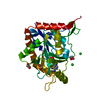
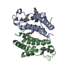
| Entry | Database: PDB / ID: 1b8n | ||||||
|---|---|---|---|---|---|---|---|
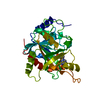
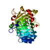
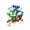
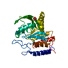
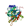
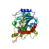
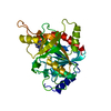
| Title | PURINE NUCLEOSIDE PHOSPHORYLASE | ||||||
 Components Components | PURINE NUCLEOSIDE PHOSPHORYLASE | ||||||
 Keywords Keywords | TRANSFERASE / PENTOSYLTRANSFERASE / PURINE NUCLEOSIDE PHOSPHORYLASE | ||||||
| Function / homology |  Function and homology information Function and homology informationguanosine phosphorylase activity / purine-nucleoside phosphorylase activity / purine-nucleoside phosphorylase / purine ribonucleoside salvage / cytosol / cytoplasm Similarity search - Function | ||||||
| Biological species |  | ||||||
| Method |  X-RAY DIFFRACTION / SYNCHROTRON / MOLECULAR REPLACEMENT / Resolution: 2 Å X-RAY DIFFRACTION / SYNCHROTRON / MOLECULAR REPLACEMENT / Resolution: 2 Å | ||||||
 Authors Authors | Fedorov, A.A. / Kicska, G.A. / Fedorov, E.V. / Strokopytov, B.V. / Tyler, P.C. / Furneaux, R.H. / Schramm, V.L. / Almo, S.C. | ||||||
 Citation Citation | Journal: Biochemistry / Year: 2002 Title: Atomic dissection of the hydrogen bond network for transition-state analogue binding to purine nucleoside phosphorylase Authors: Kicska, G.A. / Tyler, P.C. / Evans, G.B. / Furneaux, R.H. / Shi, W. / Fedorov, A.A. / Lewandowicz, A. / Cahill, S.M. / Almo, S.C. / Schramm, V.L. #1: Journal: Biochemistry / Year: 1998Title: Calf spleen purine nucleoside phosphorylase complexed with substrates and substrate analogues. Authors: Mao, C. / Cook, W.J. / Zhou, M. / Federov, A.A. / Almo, S.C. / Ealick, S.E. | ||||||
| History |
|
- Structure visualization
| Structure viewer | Molecule: MolmilJmol/JSmol |
|---|
- Downloads & links
-Download
| PDBx/mmCIF format | 1b8n.cif.gz | 73.4 KB | Display | PDBx/mmCIF format |
|---|---|---|---|---|
| PDB format | pdb1b8n.ent.gz | 52.4 KB | Display | PDB format |
| PDBx/mmJSON format | 1b8n.json.gz | Tree view | PDBx/mmJSON format | |
| Others |  Other downloads Other downloads |
-Validation report
| Arichive directory | https://data.pdbj.org/pub/pdb/validation_reports/b8/1b8nftp://data.pdbj.org/pub/pdb/validation_reports/b8/1b8n | HTTPS FTP |
|---|
-Related structure data
| Related structure data | 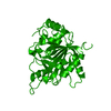 1pbnS S: Starting model for refinement |
|---|---|
| Similar structure data |

-Links
 PDBj
PDBj- Assembly
| Deposited unit | 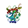
| ||||||||||
|---|---|---|---|---|---|---|---|---|---|---|---|
| 1 |
| ||||||||||
| 2 | 
| ||||||||||
| Unit cell |
|
-Components
| #1: Protein | Mass: 31687.113 Da / Num. of mol.: 1 / Source method: isolated from a natural source Details: COMPLEXED WITH TRANSITION-STATE ANALOGUE 1,4-DIDEOXY-1,4-IMINO-1-(S)-(9-DEAZAGUANIN-9-YL)-D-RIBITOL Source: (natural) References: UniProt: P55859, purine-nucleoside phosphorylase |
|---|---|
| #2: Chemical | ChemComp-MG /   Mass: 24.305 Da / Num. of mol.: 1 / Source method: obtained synthetically / Formula: Mg Mass: 24.305 Da / Num. of mol.: 1 / Source method: obtained synthetically / Formula: Mg |
| #3: Chemical | ChemComp-PO4 /   Mass: 94.971 Da / Num. of mol.: 1 / Source method: obtained synthetically / Formula: PO4 Mass: 94.971 Da / Num. of mol.: 1 / Source method: obtained synthetically / Formula: PO4 |
| #4: Chemical | ChemComp-IMG /   Mass: 281.268 Da / Num. of mol.: 1 / Source method: obtained synthetically / Formula: C11H15N5O4 Mass: 281.268 Da / Num. of mol.: 1 / Source method: obtained synthetically / Formula: C11H15N5O4 |
| #5: Water | ChemComp-HOH /  Mass: 18.015 Da / Num. of mol.: 145 / Source method: isolated from a natural source / Formula: H2O Mass: 18.015 Da / Num. of mol.: 145 / Source method: isolated from a natural source / Formula: H2O |
-Experimental details
-Experiment
| Experiment | Method: X-RAY DIFFRACTION / Number of used crystals: 1 |
|---|
- Sample preparation
| Crystal | Density Matthews: 2.22 Å3/Da / Density % sol: 43 % | ||||||||||||||||||||||||||||||||||||||||||
|---|---|---|---|---|---|---|---|---|---|---|---|---|---|---|---|---|---|---|---|---|---|---|---|---|---|---|---|---|---|---|---|---|---|---|---|---|---|---|---|---|---|---|---|
| Crystal grow | pH: 8 / Details: pH 8.0 | ||||||||||||||||||||||||||||||||||||||||||
| Components of the solutions |
| ||||||||||||||||||||||||||||||||||||||||||
| Crystal | *PLUS | ||||||||||||||||||||||||||||||||||||||||||
| Crystal grow | *PLUS Method: vapor diffusion, hanging drop / Details: Fedorov, R.H., (2001) Biochemistry, 40, 853. | ||||||||||||||||||||||||||||||||||||||||||
| Components of the solutions | *PLUS
|
-Data collection
| Diffraction | Mean temperature: 140 K |
|---|---|
| Diffraction source | Source: SYNCHROTRON / Site: NSLS  / Beamline: X9B / Wavelength: 1.04019 / Beamline: X9B / Wavelength: 1.04019 |
| Detector | Type: MARRESEARCH / Detector: IMAGE PLATE / Date: Oct 15, 1998 |
| Radiation | Protocol: SINGLE WAVELENGTH / Monochromatic (M) / Laue (L): M / Scattering type: x-ray |
| Radiation wavelength | Wavelength: 1.04019 Å / Relative weight: 1 |
| Reflection | Resolution: 2→20 Å / Num. obs: 16237 / % possible obs: 99.9 % / Redundancy: 4 % / Rmerge(I) obs: 0.08 / Net I/σ(I): 20 |
| Reflection shell | Resolution: 2→2.09 Å / Rmerge(I) obs: 0.49 / Mean I/σ(I) obs: 4.1 |
| Reflection | *PLUS % possible obs: 86.8 % / Rmerge(I) obs: 0.077 |
| Reflection shell | *PLUS % possible obs: 76.8 % |
- Processing
| Software |
| ||||||||||||||||||||||||||||||||||||||||||||||||||||||||||||
|---|---|---|---|---|---|---|---|---|---|---|---|---|---|---|---|---|---|---|---|---|---|---|---|---|---|---|---|---|---|---|---|---|---|---|---|---|---|---|---|---|---|---|---|---|---|---|---|---|---|---|---|---|---|---|---|---|---|---|---|---|---|
| Refinement | Method to determine structure: MOLECULAR REPLACEMENT Starting model: PDB ENTRY 1PBN Resolution: 2→8 Å / Cross valid method: THROUGHOUT / σ(F): 2
| ||||||||||||||||||||||||||||||||||||||||||||||||||||||||||||
| Displacement parameters | Biso mean: 26.9 Å2 | ||||||||||||||||||||||||||||||||||||||||||||||||||||||||||||
| Refinement step | Cycle: LAST / Resolution: 2→8 Å
| ||||||||||||||||||||||||||||||||||||||||||||||||||||||||||||
| Refine LS restraints |
| ||||||||||||||||||||||||||||||||||||||||||||||||||||||||||||
| LS refinement shell | Resolution: 2→2.09 Å / Total num. of bins used: 8
| ||||||||||||||||||||||||||||||||||||||||||||||||||||||||||||
| Xplor file |
| ||||||||||||||||||||||||||||||||||||||||||||||||||||||||||||
| Software | *PLUS Name: X-PLOR / Version: 3.851 / Classification: refinement | ||||||||||||||||||||||||||||||||||||||||||||||||||||||||||||
| Refinement | *PLUS σ(F): 2 / % reflection Rfree: 5 % / Rfactor obs: 0.164 | ||||||||||||||||||||||||||||||||||||||||||||||||||||||||||||
| Solvent computation | *PLUS | ||||||||||||||||||||||||||||||||||||||||||||||||||||||||||||
| Displacement parameters | *PLUS Biso mean: 26.9 Å2 | ||||||||||||||||||||||||||||||||||||||||||||||||||||||||||||
| Refine LS restraints | *PLUS
| ||||||||||||||||||||||||||||||||||||||||||||||||||||||||||||
| LS refinement shell | *PLUS Rfactor Rfree: 0.296 / % reflection Rfree: 5 % / Rfactor Rwork: 0.241 |
