 Movie
Movie Controller


+ Open data

- Basic information
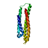
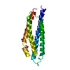
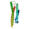
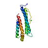
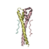
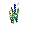
| Entry | Database: PDB / ID: 1b68 | ||||||
|---|---|---|---|---|---|---|---|
| Title | APOLIPOPROTEIN E4 (APOE4), 22K FRAGMENT | ||||||
 Components Components | APOLIPOPROTEIN E | ||||||
 Keywords Keywords | LIPID TRANSPORT / HEPARIN-BINDING / PLASMA PROTEIN / HDL / VLDL | ||||||
| Function / homology |  Function and homology information Function and homology informationlipid transport involved in lipid storage / maintenance of location in cell / intermediate-density lipoprotein particle clearance / positive regulation of lipid transport across blood-brain barrier / regulation of cellular response to very-low-density lipoprotein particle stimulus / metal chelating activity / triglyceride-rich lipoprotein particle clearance / negative regulation of triglyceride metabolic process / discoidal high-density lipoprotein particle / Transcriptional regulation by the AP-2 (TFAP2) family of transcription factors ...lipid transport involved in lipid storage / maintenance of location in cell / intermediate-density lipoprotein particle clearance / positive regulation of lipid transport across blood-brain barrier / regulation of cellular response to very-low-density lipoprotein particle stimulus / metal chelating activity / triglyceride-rich lipoprotein particle clearance / negative regulation of triglyceride metabolic process / discoidal high-density lipoprotein particle / Transcriptional regulation by the AP-2 (TFAP2) family of transcription factors / chylomicron remnant clearance / chylomicron remnant / lipoprotein particle / negative regulation of cholesterol biosynthetic process / regulation of amyloid-beta clearance / intermediate-density lipoprotein particle / NMDA glutamate receptor clustering / very-low-density lipoprotein particle remodeling / Chylomicron clearance / acylglycerol homeostasis / phosphatidylcholine-sterol O-acyltransferase activator activity / positive regulation of phospholipid efflux / Chylomicron remodeling / lipid carrier activity / positive regulation of low-density lipoprotein particle receptor catabolic process / cellular response to lipoprotein particle stimulus / very-low-density lipoprotein particle clearance / Chylomicron assembly / high-density lipoprotein particle clearance / phospholipid efflux / lipoprotein catabolic process / very-low-density lipoprotein particle receptor binding / regulation of protein metabolic process / chylomicron / high-density lipoprotein particle remodeling / lipoprotein biosynthetic process / multivesicular body, internal vesicle / positive regulation of amyloid-beta clearance / reverse cholesterol transport / positive regulation of cholesterol metabolic process / response to caloric restriction / regulation of amyloid fibril formation / high-density lipoprotein particle assembly / host-mediated activation of viral process / melanosome organization / cholesterol transfer activity / low-density lipoprotein particle / cholesterol catabolic process / protein import / high-density lipoprotein particle / very-low-density lipoprotein particle / low-density lipoprotein particle remodeling / amyloid precursor protein metabolic process / regulation of behavioral fear response / negative regulation of amyloid fibril formation / triglyceride metabolic process / heparan sulfate proteoglycan binding / positive regulation of membrane protein ectodomain proteolysis / negative regulation of endothelial cell migration / regulation of Cdc42 protein signal transduction / regulation of amyloid precursor protein catabolic process / HDL remodeling / negative regulation of protein metabolic process / artery morphogenesis / cholesterol efflux / regulation of cholesterol metabolic process / regulation of axon extension / triglyceride homeostasis / Scavenging by Class A Receptors / low-density lipoprotein particle receptor binding / positive regulation of amyloid fibril formation / virion assembly / synaptic transmission, cholinergic / regulation of innate immune response / positive regulation of dendritic spine development / negative regulation of endothelial cell proliferation / antioxidant activity / negative regulation of platelet-derived growth factor receptor signaling pathway / positive regulation of lipoprotein transport / response to dietary excess / negative regulation of amyloid-beta formation / lipoprotein particle binding / AMPA glutamate receptor clustering / negative regulation of blood vessel endothelial cell migration / negative regulation of long-term synaptic potentiation / negative regulation of platelet activation / locomotory exploration behavior / regulation of neuronal synaptic plasticity / negative regulation of blood coagulation / positive regulation of cholesterol efflux / negative regulation of protein secretion / positive regulation of dendritic spine maintenance / fatty acid homeostasis / intracellular transport / regulation of protein-containing complex assembly / positive regulation of endocytosis / long-chain fatty acid transport / Nuclear signaling by ERBB4 / positive regulation of lipid biosynthetic process / cholesterol metabolic process Similarity search - Function | ||||||
| Biological species |  Homo sapiens (human) Homo sapiens (human) | ||||||
| Method |  X-RAY DIFFRACTION / SYNCHROTRON / MOLECULAR REPLACEMENT / Resolution: 2 Å X-RAY DIFFRACTION / SYNCHROTRON / MOLECULAR REPLACEMENT / Resolution: 2 Å | ||||||
 Authors Authors | Rupp, B. / Peters-Libeu, C. | ||||||
 Citation Citation | Journal: Biochemistry / Year: 2001 Title: Interaction of the N-terminal domain of apolipoprotein E4 with heparin. Authors: Dong, J. / Peters-Libeu, C.A. / Weisgraber, K.H. / Segelke, B.W. / Rupp, B. / Capila, I. / Hernaiz, M.J. / LeBrun, L.A. / Linhardt, R.J. | ||||||
| History |
|
- Structure visualization
| Structure viewer | Molecule: MolmilJmol/JSmol |
|---|
- Downloads & links
-Download
| PDBx/mmCIF format | 1b68.cif.gz | 44.6 KB | Display | PDBx/mmCIF format |
|---|---|---|---|---|
| PDB format | pdb1b68.ent.gz | 30.6 KB | Display | PDB format |
| PDBx/mmJSON format | 1b68.json.gz | Tree view | PDBx/mmJSON format | |
| Others |  Other downloads Other downloads |
-Validation report
| Arichive directory | https://data.pdbj.org/pub/pdb/validation_reports/b6/1b68ftp://data.pdbj.org/pub/pdb/validation_reports/b6/1b68 | HTTPS FTP |
|---|
-Related structure data
| Related structure data |  1bz4S S: Starting model for refinement |
|---|---|
| Similar structure data |


-Links
 PDBj
PDBj


- Assembly
| Deposited unit | 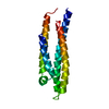
| ||||||||
|---|---|---|---|---|---|---|---|---|---|
| 1 |
| ||||||||
| Unit cell |
|
-Components
| #1: Protein | Mass: 22216.135 Da / Num. of mol.: 1 Fragment: 22K FRAGMENT, ISOFORM E4, RECEPTOR BINDING DOMAIN, RESIDUES 1-191 Source method: isolated from a genetically manipulated source Source: (gene. exp.) Homo sapiens (human) / Plasmid: PET / Species (production host): Escherichia coli / Production host:  |
|---|---|
| #2: Water | ChemComp-HOH /  Mass: 18.015 Da / Num. of mol.: 133 / Source method: isolated from a natural source / Formula: H2O Mass: 18.015 Da / Num. of mol.: 133 / Source method: isolated from a natural source / Formula: H2O |
-Experimental details
-Experiment
| Experiment | Method: X-RAY DIFFRACTION / Number of used crystals: 1 |
|---|
- Sample preparation
| Crystal | Density Matthews: 2.18 Å3/Da / Density % sol: 43.6 % Description: RIGID BODY REFINEMENT ONLY, THEN REBUILD, WARP MAPS USED | ||||||||||||||||||||
|---|---|---|---|---|---|---|---|---|---|---|---|---|---|---|---|---|---|---|---|---|---|
| Crystal grow | Temperature: 298 K / Method: vapor diffusion, hanging drop / pH: 6 Details: WELL : 28% PEG 400, 20MM NAOAC, PH 6.0, 0.1% BME PROTEIN SOLN: 40MM (NH4)H(CO3), 7MG/ML PROTEIN DROPS : WELL/PROTEIN 1/3, ROOM TEMPERATURE, pH 6.00, VAPOR DIFFUSION, HANGING DROP, temperature 298K | ||||||||||||||||||||
| Crystal grow | *PLUS pH: 6.2 | ||||||||||||||||||||
| Components of the solutions | *PLUS
|
-Data collection
| Diffraction | Mean temperature: 125 K |
|---|---|
| Diffraction source | Source: SYNCHROTRON / Site: ALS  / Beamline: 5.0.1 / Wavelength: 1 / Beamline: 5.0.1 / Wavelength: 1 |
| Detector | Type: ADSC / Detector: CCD / Date: Mar 1, 1998 |
| Radiation | Protocol: SINGLE WAVELENGTH / Monochromatic (M) / Laue (L): M / Scattering type: x-ray |
| Radiation wavelength | Wavelength: 1 Å / Relative weight: 1 |
| Reflection | Resolution: 1.84→25 Å / Num. obs: 16493 / % possible obs: 99.1 % / Observed criterion σ(I): 0 / Redundancy: 4.9 % / Biso Wilson estimate: 29.2 Å2 / Rmerge(I) obs: 0.037 / Rsym value: 0.037 / Net I/σ(I): 27.4 |
| Reflection shell | Resolution: 1.84→1.98 Å / Redundancy: 3.1 % / Rmerge(I) obs: 0.199 / Mean I/σ(I) obs: 3.7 / Rsym value: 0.199 / % possible all: 98.7 |
| Reflection shell | *PLUS % possible obs: 98.7 % |
- Processing
| Software |
| ||||||||||||||||||||||||||||||||||||||||||||||||||||||||||||
|---|---|---|---|---|---|---|---|---|---|---|---|---|---|---|---|---|---|---|---|---|---|---|---|---|---|---|---|---|---|---|---|---|---|---|---|---|---|---|---|---|---|---|---|---|---|---|---|---|---|---|---|---|---|---|---|---|---|---|---|---|---|
| Refinement | Method to determine structure: MOLECULAR REPLACEMENT Starting model: 1BZ4 Resolution: 2→8 Å / Rfactor Rfree error: 0.007 / Data cutoff high absF: 10000000 / Data cutoff low absF: 0.001 / Isotropic thermal model: RESTRAINED / Cross valid method: THROUGHOUT / σ(F): 2 / Details: BULK SOLVENT MODEL USED
| ||||||||||||||||||||||||||||||||||||||||||||||||||||||||||||
| Displacement parameters | Biso mean: 38.7 Å2
| ||||||||||||||||||||||||||||||||||||||||||||||||||||||||||||
| Refine analyze |
| ||||||||||||||||||||||||||||||||||||||||||||||||||||||||||||
| Refinement step | Cycle: LAST / Resolution: 2→8 Å
| ||||||||||||||||||||||||||||||||||||||||||||||||||||||||||||
| Refine LS restraints |
| ||||||||||||||||||||||||||||||||||||||||||||||||||||||||||||
| LS refinement shell | Resolution: 2→2.12 Å / Rfactor Rfree error: 0.024 / Total num. of bins used: 6
| ||||||||||||||||||||||||||||||||||||||||||||||||||||||||||||
| Xplor file |
| ||||||||||||||||||||||||||||||||||||||||||||||||||||||||||||
| Software | *PLUS Name: X-PLOR / Version: 3.851 / Classification: refinement | ||||||||||||||||||||||||||||||||||||||||||||||||||||||||||||
| Refinement | *PLUS Rfactor obs: 0.2 / Rfactor Rwork: 0.2 | ||||||||||||||||||||||||||||||||||||||||||||||||||||||||||||
| Solvent computation | *PLUS | ||||||||||||||||||||||||||||||||||||||||||||||||||||||||||||
| Displacement parameters | *PLUS | ||||||||||||||||||||||||||||||||||||||||||||||||||||||||||||
| Refine LS restraints | *PLUS
| ||||||||||||||||||||||||||||||||||||||||||||||||||||||||||||
| LS refinement shell | *PLUS Rfactor Rfree: 0.321 / Rfactor Rwork: 0.298 |
