 Movie
Movie Controller

[English] 日本語
 Yorodumi
- PDB-1azc: STRUCTURE OF APO-AZURIN FROM ALCALIGENES DENITRIFICANS AT 1.8 ANG... -
+ Open data

- Basic information
| Entry | Database: PDB / ID: 1azc | ||||||
|---|---|---|---|---|---|---|---|
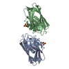
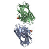
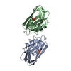
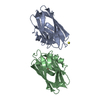
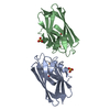
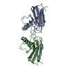
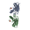
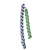
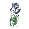
| Title | STRUCTURE OF APO-AZURIN FROM ALCALIGENES DENITRIFICANS AT 1.8 ANGSTROMS RESOLUTION | ||||||
 Components Components | AZURIN | ||||||
 Keywords Keywords | ELECTRON TRANSPORT(COPPER) | ||||||
| Function / homology |  Function and homology information Function and homology information | ||||||
| Biological species |  Achromobacter xylosoxidans (bacteria) Achromobacter xylosoxidans (bacteria) | ||||||
| Method |  X-RAY DIFFRACTION / Resolution: 1.8 Å X-RAY DIFFRACTION / Resolution: 1.8 Å | ||||||
 Authors Authors | Baker, E.N. / Shepard, W.E.B. / Kingston, R.L. | ||||||
 Citation Citation | Journal: Acta Crystallogr.,Sect.D / Year: 1993 Title: Structure of apo-azurin from Alcaligenes denitrificans at 1.8 A resolution. Authors: Shepard, W.E. / Kingston, R.L. / Anderson, B.F. / Baker, E.N. #1: Journal: J.Am.Chem.Soc. / Year: 1990Title: Copper Coordination Geometry in Azurin Undergoes Minimal Change on Reduction of Copper(II) to Copper (I) Authors: Shepard, W.E.B. / Anderson, B.F. / Lewandoski, D.A. / Norris, G.E. / Baker, E.N. #2: Journal: J.Mol.Biol. / Year: 1988Title: Structure of Azurin from Alcaligenes Denitrificans Refinement at 1.8 Angstroms Resolution and Comparison of the Two Crystallographically Independent Molecules Authors: Baker, E.N. #3: Journal: J.Mol.Biol. / Year: 1983Title: Structure of Azurin from Alcaligenes Denitrificans at 2.5 Angstroms Resolution Authors: Norris, G.E. / Anderson, B.F. / Baker, E.N. #4: Journal: J.Mol.Biol. / Year: 1979Title: Purification and Preliminary Crystallographic Studies on Azurin and Cytochrome C' from Alcaligenes Denitrificans and Alcaligenes Sp. Ncib 11015 Authors: Norris, G.E. / Anderson, B.F. / Baker, E.N. / Rumball, S.V. | ||||||
| History |
|
- Structure visualization
| Structure viewer | Molecule: MolmilJmol/JSmol |
|---|
- Downloads & links
-Download
| PDBx/mmCIF format | 1azc.cif.gz | 68.2 KB | Display | PDBx/mmCIF format |
|---|---|---|---|---|
| PDB format | pdb1azc.ent.gz | 50.4 KB | Display | PDB format |
| PDBx/mmJSON format | 1azc.json.gz | Tree view | PDBx/mmJSON format | |
| Others |  Other downloads Other downloads |
-Validation report
| Arichive directory | https://data.pdbj.org/pub/pdb/validation_reports/az/1azcftp://data.pdbj.org/pub/pdb/validation_reports/az/1azc | HTTPS FTP |
|---|
-Related structure data


-Links
 PDBj
PDBj- Assembly
| Deposited unit | 
| ||||||||
|---|---|---|---|---|---|---|---|---|---|
| 1 |
| ||||||||
| Unit cell |
| ||||||||
| Noncrystallographic symmetry (NCS) | NCS oper: (Code: given Matrix: (-0.098, 0.995, 0.018), Vector: Details | THE ASYMMETRIC UNIT CONTAINS TWO CRYSTALLOGRAPHICALLY INDEPENDENT MOLECULES, A AND B, RELATED BY AN APPROXIMATE TWO-FOLD AXIS. THE TRANSFORMATION PRESENTED ON *MTRIX* RECORDS BELOW WILL YIELD APPROXIMATE COORDINATES FOR MOLECULE A WHEN APPLIED TO MOLECULE B. | |
-Components
| #1: Protein | Mass: 14008.003 Da / Num. of mol.: 2 Source method: isolated from a genetically manipulated source Source: (gene. exp.) Achromobacter xylosoxidans (bacteria) / References: UniProt: P00280#2: Chemical |   Mass: 63.546 Da / Num. of mol.: 2 / Source method: obtained synthetically / Formula: Cu Mass: 63.546 Da / Num. of mol.: 2 / Source method: obtained synthetically / Formula: Cu#3: Chemical | ChemComp-SO4 /   Mass: 96.063 Da / Num. of mol.: 4 / Source method: obtained synthetically / Formula: SO4 Mass: 96.063 Da / Num. of mol.: 4 / Source method: obtained synthetically / Formula: SO4#4: Water | ChemComp-HOH / |  Mass: 18.015 Da / Num. of mol.: 246 / Source method: isolated from a natural source / Formula: H2O Mass: 18.015 Da / Num. of mol.: 246 / Source method: isolated from a natural source / Formula: H2OHas protein modification | Y | Sequence details | AMINO ACID SEQUENCE IS AS IN C.W.G. HOITINK, L.P. WOUDT, J.C.M. TURENHOUT, M. VAN DE KAMP, G.W. ...AMINO ACID SEQUENCE IS AS IN C.W.G. HOITINK, L.P. WOUDT, J.C.M. TURENHOUT, M. VAN DE KAMP, G.W. CARTERS (1990) GENE 90, 15-20. | |
|---|
-Experimental details
-Experiment
| Experiment | Method: X-RAY DIFFRACTION |
|---|
- Sample preparation
| Crystal | Density Matthews: 2.47 Å3/Da / Density % sol: 50.21 % | |||||||||||||||||||||||||
|---|---|---|---|---|---|---|---|---|---|---|---|---|---|---|---|---|---|---|---|---|---|---|---|---|---|---|
| Crystal grow | *PLUS pH: 6 / Method: interface diffusion | |||||||||||||||||||||||||
| Components of the solutions | *PLUS
|
-Data collection
| Radiation | Scattering type: x-ray |
|---|---|
| Radiation wavelength | Relative weight: 1 |
| Reflection | *PLUS Highest resolution: 1.8 Å / Num. obs: 24722 / % possible obs: 89 % / Observed criterion σ(I): 2 / Rmerge(I) obs: 0.055 |
- Processing
| Software | Name: PROLSQ / Classification: refinement | |||||||||||||||||||||||||||||||||||||||||||||||||||||||||||||||
|---|---|---|---|---|---|---|---|---|---|---|---|---|---|---|---|---|---|---|---|---|---|---|---|---|---|---|---|---|---|---|---|---|---|---|---|---|---|---|---|---|---|---|---|---|---|---|---|---|---|---|---|---|---|---|---|---|---|---|---|---|---|---|---|---|
| Refinement | Resolution: 1.8→10 Å / σ(F): 0 /
| |||||||||||||||||||||||||||||||||||||||||||||||||||||||||||||||
| Refinement step | Cycle: LAST / Resolution: 1.8→10 Å
| |||||||||||||||||||||||||||||||||||||||||||||||||||||||||||||||
| Refine LS restraints |
| |||||||||||||||||||||||||||||||||||||||||||||||||||||||||||||||
| Refinement | *PLUS Highest resolution: 1.8 Å / Lowest resolution: 10 Å / Num. reflection all: 22667 / σ(F): 0 / Rfactor all: 0.16 | |||||||||||||||||||||||||||||||||||||||||||||||||||||||||||||||
| Solvent computation | *PLUS | |||||||||||||||||||||||||||||||||||||||||||||||||||||||||||||||
| Displacement parameters | *PLUS | |||||||||||||||||||||||||||||||||||||||||||||||||||||||||||||||
| Refine LS restraints | *PLUS Type: p_angle_d / Dev ideal: 0.045 |
