 Movie
Movie Controller

[English] 日本語
 Yorodumi
- PDB-1am5: THE CRYSTAL STRUCTURE AND PROPOSED AMINO ACID SEQUENCE OF A PEPSI... -
+ Open data

- Basic information
| Entry | Database: PDB / ID: 1am5 | ||||||
|---|---|---|---|---|---|---|---|
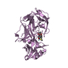
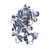
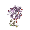
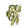
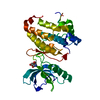
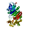
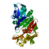
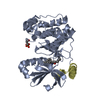
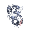
| Title | THE CRYSTAL STRUCTURE AND PROPOSED AMINO ACID SEQUENCE OF A PEPSIN FROM ATLANTIC COD (GADUS MORHUA) | ||||||
 Components Components | PEPSIN | ||||||
 Keywords Keywords | ASPARTYL PROTEASE / ACID PROTEINASE / HYDROLASE | ||||||
| Function / homology |  Function and homology information Function and homology informationHydrolases; Acting on peptide bonds (peptidases); Aspartic endopeptidases / digestion / aspartic-type endopeptidase activity / proteolysis Similarity search - Function | ||||||
| Biological species |  Gadus morhua (Atlantic cod) Gadus morhua (Atlantic cod) | ||||||
| Method |  X-RAY DIFFRACTION / MOLECULAR REPLACEMENT / Resolution: 2.16 Å X-RAY DIFFRACTION / MOLECULAR REPLACEMENT / Resolution: 2.16 Å | ||||||
 Authors Authors | Karlsen, S. / Hough, E. / Olsen, R.L. | ||||||
 Citation Citation | Journal: Acta Crystallogr.,Sect.D / Year: 1998 Title: Structure and proposed amino-acid sequence of a pepsin from atlantic cod (Gadus morhua). Authors: Karlsen, S. / Hough, E. / Olsen, R.L. #1: Journal: COMP.BIOCHEM.PHYSIOL. B: BIOCHEM.MOL.BIOL. / Year: 1990Title: Catalytic Properties and Chemical Composition of Pepsins from Atlantic Cod (Gadus Morhua) Authors: Gildberg, A. / Olsen, R.L. / Bjarnason, J.B. | ||||||
| History |
|
- Structure visualization
| Structure viewer | Molecule: MolmilJmol/JSmol |
|---|
- Downloads & links
-Download
| PDBx/mmCIF format | 1am5.cif.gz | 79 KB | Display | PDBx/mmCIF format |
|---|---|---|---|---|
| PDB format | pdb1am5.ent.gz | 56.7 KB | Display | PDB format |
| PDBx/mmJSON format | 1am5.json.gz | Tree view | PDBx/mmJSON format | |
| Others |  Other downloads Other downloads |
-Validation report
| Arichive directory | https://data.pdbj.org/pub/pdb/validation_reports/am/1am5ftp://data.pdbj.org/pub/pdb/validation_reports/am/1am5 | HTTPS FTP |
|---|
-Related structure data
| Related structure data |  5pepS S: Starting model for refinement |
|---|---|
| Similar structure data |

-Links
 PDBj
PDBj- Assembly
| Deposited unit | 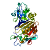
| ||||||||
|---|---|---|---|---|---|---|---|---|---|
| 1 |
| ||||||||
| Unit cell |
|
-Components
| #1: Protein | Mass: 34033.559 Da / Num. of mol.: 1 / Source method: isolated from a natural source / Source: (natural) Gadus morhua (Atlantic cod) / Organ: STOMACH / Tissue: GASTRIC MUCOSA / References: UniProt: P56272, pepsin A |
|---|---|
| #2: Water | ChemComp-HOH /  Mass: 18.015 Da / Num. of mol.: 161 / Source method: isolated from a natural source / Formula: H2O Mass: 18.015 Da / Num. of mol.: 161 / Source method: isolated from a natural source / Formula: H2O |
| Has protein modification | Y |
| Sequence details | THE AMINO ACID NUMBERING IS BASED ON THE SEQUENCE OF PEPSIN FROM PORCINE. AS RESIDUE 207 AND 241 ...THE AMINO ACID NUMBERING IS BASED ON THE SEQUENCE OF PEPSIN FROM PORCINE. AS RESIDUE 207 AND 241 NOT ARE PRESENT IN THE COD PEPSIN, THESE RESIDUES ARE DELETED IN THE COORDINATE |
-Experimental details
-Experiment
| Experiment | Method: X-RAY DIFFRACTION / Number of used crystals: 1 |
|---|
- Sample preparation
| Crystal | Density Matthews: 2.15 Å3/Da / Density % sol: 43 % | ||||||||||||||||||||||||||||||
|---|---|---|---|---|---|---|---|---|---|---|---|---|---|---|---|---|---|---|---|---|---|---|---|---|---|---|---|---|---|---|---|
| Crystal grow | pH: 5.4 / Details: 7.5 % 2-PROPANOL, 100 MM SODIUM ACETATE PH 5.4 | ||||||||||||||||||||||||||||||
| Crystal grow | *PLUS Temperature: 277 K / Method: vapor diffusion, hanging drop | ||||||||||||||||||||||||||||||
| Components of the solutions | *PLUS
|
-Data collection
| Diffraction | Mean temperature: 291 K |
|---|---|
| Diffraction source | Type: BRUKER NONIUS / Wavelength: 1.5418 |
| Detector | Type: ENRAF-NONIUS FAST / Detector: DIFFRACTOMETER / Date: Oct 1, 1995 / Details: COLLIMATOR |
| Radiation | Monochromator: GRAPHITE(002) / Monochromatic (M) / Laue (L): M / Scattering type: x-ray |
| Radiation wavelength | Wavelength: 1.5418 Å / Relative weight: 1 |
| Reflection | Resolution: 2.16→20 Å / Num. obs: 13687 / % possible obs: 83.7 % / Observed criterion σ(I): 1 / Redundancy: 2.7 % / Biso Wilson estimate: 22.3 Å2 / Rsym value: 0.066 / Net I/σ(I): 10.8 |
| Reflection shell | Resolution: 2.16→2.28 Å / Redundancy: 1.4 % / Mean I/σ(I) obs: 3.5 / Rsym value: 0.221 / % possible all: 41.4 |
| Reflection | *PLUS Num. measured all: 37108 / Rmerge(I) obs: 0.066 |
| Reflection shell | *PLUS % possible obs: 41.4 % / Num. unique obs: 948 / Num. measured obs: 1322 / Rmerge(I) obs: 0.221 |
- Processing
| Software |
| |||||||||||||||||||||||||||||||||||||||||||||||||||||||||||||||
|---|---|---|---|---|---|---|---|---|---|---|---|---|---|---|---|---|---|---|---|---|---|---|---|---|---|---|---|---|---|---|---|---|---|---|---|---|---|---|---|---|---|---|---|---|---|---|---|---|---|---|---|---|---|---|---|---|---|---|---|---|---|---|---|---|
| Refinement | Method to determine structure: MOLECULAR REPLACEMENT Starting model: PDB ENTRY 5PEP Resolution: 2.16→8 Å / σ(F): 3
| |||||||||||||||||||||||||||||||||||||||||||||||||||||||||||||||
| Displacement parameters | Biso mean: 17.22 Å2 | |||||||||||||||||||||||||||||||||||||||||||||||||||||||||||||||
| Refine analyze | Luzzati coordinate error obs: 0.2 Å | |||||||||||||||||||||||||||||||||||||||||||||||||||||||||||||||
| Refinement step | Cycle: LAST / Resolution: 2.16→8 Å
| |||||||||||||||||||||||||||||||||||||||||||||||||||||||||||||||
| Refine LS restraints |
| |||||||||||||||||||||||||||||||||||||||||||||||||||||||||||||||
| Software | *PLUS Name: PROLSQ / Classification: refinement | |||||||||||||||||||||||||||||||||||||||||||||||||||||||||||||||
| Refinement | *PLUS Rfactor obs: 0.208 | |||||||||||||||||||||||||||||||||||||||||||||||||||||||||||||||
| Solvent computation | *PLUS | |||||||||||||||||||||||||||||||||||||||||||||||||||||||||||||||
| Displacement parameters | *PLUS |
