 Movie
Movie Controller

[English] 日本語
 Yorodumi
- PDB-4pep: THE MOLECULAR AND CRYSTAL STRUCTURES OF MONOCLINIC PORCINE PEPSIN... -
+ Open data

- Basic information
| Entry | Database: PDB / ID: 4pep | |||||||||
|---|---|---|---|---|---|---|---|---|---|---|
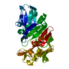
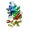
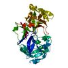
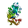
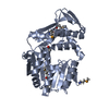
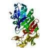
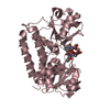
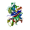
| Title | THE MOLECULAR AND CRYSTAL STRUCTURES OF MONOCLINIC PORCINE PEPSIN REFINED AT 1.8 ANGSTROMS RESOLUTION | |||||||||
 Components Components | PEPSIN | |||||||||
 Keywords Keywords | HYDROLASE (ACID PROTEINASE) | |||||||||
| Function / homology |  Function and homology information Function and homology informationSurfactant metabolism / pepsin A / digestion / aspartic-type endopeptidase activity / proteolysis / extracellular region Similarity search - Function | |||||||||
| Biological species |  | |||||||||
| Method |  X-RAY DIFFRACTION / Resolution: 1.8 Å X-RAY DIFFRACTION / Resolution: 1.8 Å | |||||||||
 Authors Authors | Andreeva, N. / Fedorov, A.A. / Sielecki, A. / James, M. | |||||||||
 Citation Citation | Journal: J.Mol.Biol. / Year: 1990 Title: Molecular and crystal structures of monoclinic porcine pepsin refined at 1.8 A resolution. Authors: Sielecki, A.R. / Fedorov, A.A. / Boodhoo, A. / Andreeva, N.S. / James, M.N. | |||||||||
| History |
| |||||||||
| Remark 700 | SHEET THE SHEET IDENTIFIED AS *I* BY THE DEPOSITORS CONTAINS A BIFURCATED STRAND. THIS IS ...SHEET THE SHEET IDENTIFIED AS *I* BY THE DEPOSITORS CONTAINS A BIFURCATED STRAND. THIS IS REPRESENTED BY DEFINING THE SHEET TWICE ON *SHEET* RECORDS BELOW. THUS SHEETS *IA* *IB* DIFFER ONLY IN STRAND 6. |
- Structure visualization
| Structure viewer | Molecule: MolmilJmol/JSmol |
|---|
- Downloads & links
-Download
| PDBx/mmCIF format | 4pep.cif.gz | 78.5 KB | Display | PDBx/mmCIF format |
|---|---|---|---|---|
| PDB format | pdb4pep.ent.gz | 57.3 KB | Display | PDB format |
| PDBx/mmJSON format | 4pep.json.gz | Tree view | PDBx/mmJSON format | |
| Others |  Other downloads Other downloads |
-Validation report
| Arichive directory | https://data.pdbj.org/pub/pdb/validation_reports/pe/4pepftp://data.pdbj.org/pub/pdb/validation_reports/pe/4pep | HTTPS FTP |
|---|
-Related structure data
| Similar structure data |
|---|


-Links
 PDBj
PDBj
- Assembly
| Deposited unit | 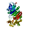
| ||||||||
|---|---|---|---|---|---|---|---|---|---|
| 1 |
| ||||||||
| Unit cell |
| ||||||||
| Atom site foot note | 1: RESIDUE PRO 23 IS A CIS PROLINE. 2: AS DETAILED IN THE REFERENCE CITED ON THE *JRNL* RECORDS ABOVE, THE FOLLOWING LOOPS ARE POORLY DETERMINED IN THE STRUCTURE PRESENTED IN THIS ENTRY (B FACTORS .GT. 40A**2) -ASP 200 - THR 203 SER ...2: AS DETAILED IN THE REFERENCE CITED ON THE *JRNL* RECORDS ABOVE, THE FOLLOWING LOOPS ARE POORLY DETERMINED IN THE STRUCTURE PRESENTED IN THIS ENTRY (B FACTORS .GT. 40A**2) -ASP 200 - THR 203 SER 241 - GLY 243 SER 250 - ASP 253 GLN 277 - SER 281 ASP 290 - GLU 297 IN ADDITION, THE SIDE CHAINS OF RESIDUES LEU 166 AND GLU 244 - ILE 247 HAVE POORLY DEFINED ELECTRON DENSITY AND/OR COULD HAVE MORE THAN ONE POSSIBLE CONFORMATION. |
-Components
| #1: Protein | Mass: 34594.469 Da / Num. of mol.: 1 Source method: isolated from a genetically manipulated source Source: (gene. exp.) |
|---|---|
| #2: Water | ChemComp-HOH /  Mass: 18.015 Da / Num. of mol.: 187 / Source method: isolated from a natural source / Formula: H2O Mass: 18.015 Da / Num. of mol.: 187 / Source method: isolated from a natural source / Formula: H2O |
| Has protein modification | Y |
-Experimental details
-Experiment
| Experiment | Method: X-RAY DIFFRACTION |
|---|
- Sample preparation
| Crystal | Density Matthews: 2.07 Å3/Da / Density % sol: 40.47 % | |||||||||||||||
|---|---|---|---|---|---|---|---|---|---|---|---|---|---|---|---|---|
| Crystal grow | *PLUS pH: 2 / Method: other / Details: pH is adjusted to 2.0 with 1M H2SO4 | |||||||||||||||
| Components of the solutions | *PLUS
|
-Data collection
| Radiation | Scattering type: x-ray |
|---|---|
| Radiation wavelength | Relative weight: 1 |
| Reflection | *PLUS Highest resolution: 1.8 Å / Num. obs: 23897 / Num. measured all: 90730 / Rmerge(I) obs: 0.062 |
- Processing
| Software | Name: PROLSQ / Classification: refinement | ||||||||||||||||||||||||||||||||||||||||||||||||||||||||||||||||||||||||||||||||||||
|---|---|---|---|---|---|---|---|---|---|---|---|---|---|---|---|---|---|---|---|---|---|---|---|---|---|---|---|---|---|---|---|---|---|---|---|---|---|---|---|---|---|---|---|---|---|---|---|---|---|---|---|---|---|---|---|---|---|---|---|---|---|---|---|---|---|---|---|---|---|---|---|---|---|---|---|---|---|---|---|---|---|---|---|---|---|
| Refinement | Resolution: 1.8→8 Å / σ(F): 3 Details: 187 SOLVENTS ARE INCLUDED IN THIS ENTRY (HOH 328 TO 514). THOSE WITH LOWER RESIDUE NUMBERS (LOWER B FACTORS, HIGHER OCCUPANCIES) ARE MORE RELIABLE THAN THOSE LOCATED TOWARDS THE END OF THE ...Details: 187 SOLVENTS ARE INCLUDED IN THIS ENTRY (HOH 328 TO 514). THOSE WITH LOWER RESIDUE NUMBERS (LOWER B FACTORS, HIGHER OCCUPANCIES) ARE MORE RELIABLE THAN THOSE LOCATED TOWARDS THE END OF THE LIST (HIGHER B FACTORS, LOWER OCCUPANCIES).
| ||||||||||||||||||||||||||||||||||||||||||||||||||||||||||||||||||||||||||||||||||||
| Refinement step | Cycle: LAST / Resolution: 1.8→8 Å
| ||||||||||||||||||||||||||||||||||||||||||||||||||||||||||||||||||||||||||||||||||||
| Refine LS restraints |
| ||||||||||||||||||||||||||||||||||||||||||||||||||||||||||||||||||||||||||||||||||||
| Refinement | *PLUS σ(F): 3 / Highest resolution: 1.8 Å / Lowest resolution: 8 Å / Num. reflection obs: 20519 / Rfactor obs: 0.174 | ||||||||||||||||||||||||||||||||||||||||||||||||||||||||||||||||||||||||||||||||||||
| Solvent computation | *PLUS | ||||||||||||||||||||||||||||||||||||||||||||||||||||||||||||||||||||||||||||||||||||
| Displacement parameters | *PLUS |
