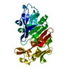
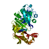
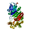
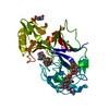
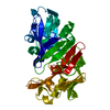
SHEET SHEET VI CORRESPONDS TO SHEET C2 (POSSIBLY FOR PSI2) OF PROTEIN DATA BANK ENTRY *2APR* ...SHEET SHEET VI CORRESPONDS TO SHEET C2 (POSSIBLY FOR PSI2) OF PROTEIN DATA BANK ENTRY *2APR* (RHIZOPUSPEPSIN, A SIMILAR ASPARTYL PROTEINASE). IN PEPSIN THIS SHEET HAS ONE MORE STRAND. THE EXTENDED SHEET VII CORRESPONDS TO SHEETS C1 (POSSIBLY BECAUSE OF PSI1) AND BIFURCATED SHEET 4A OF *2APR**. THE EXTENDED SHEET DESCRIPTION WAS USED IN THIS ENTRY BECAUSE THERE ARE THREE GOOD HYDROGEN BONDS BETWEEN STRANDS 3 AND 4. THE OVERALL STRUCTURE IS WELL CONSERVED BETWEEN THE TWO ENZYMES AS WELL AS AMONG OTHER FUNGAL PROTEINASES (PENNICILLOPEPSIN AND ENDOTHIAPEPSIN).
1: THE ELECTRON DENSITY FOR RESIDUES 292 - 296 IS WEAK IN THE FINAL MAP, INDICATING HIGH MOBILITY AND/OR DISORDER. THE CONFORMATION FOR THESE RESIDUES SHOULD BE CONSIDERED AS TENTATIVE. THE ELECTRON ...1: THE ELECTRON DENSITY FOR RESIDUES 292 - 296 IS WEAK IN THE FINAL MAP, INDICATING HIGH MOBILITY AND/OR DISORDER. THE CONFORMATION FOR THESE RESIDUES SHOULD BE CONSIDERED AS TENTATIVE. THE ELECTRON DENSITY IS ALSO WEAK FOR RESIDUES IN THE TURN ASP 278 - SER 281. 2: RESIDUE PRO 23 IS A CIS PROLINE.
-
Components
#1: Protein
PEPSIN
Mass: 34514.492 Da / Num. of mol.: 1 Source method: isolated from a genetically manipulated source Source: (gene. exp.) Sus scrofa (pig) / References: UniProt: P00791, pepsin A
Mass: 18.015 Da / Num. of mol.: 206 / Source method: isolated from a natural source / Formula: H2O
Has protein modification
Y
-
Experimental details
-
Experiment
Experiment
Method: X-RAY DIFFRACTION
-
Sample preparation
Crystal
Density Matthews: 2.09 Å3/Da / Density % sol: 41.27 %
Crystal grow
*PLUS
pH: 2 / Method: unknown Details: Andreeva, N.S. et al (1984). J. Biol. Chem., 259, 11353-11365.
Components of the solutions
*PLUS
ID
Conc.
Common name
Crystal-ID
Sol-ID
1
20 %
enzyme
1
1
2
20 %
ethanol
1
1
3
2.5M
sulfuricacid
1
1
-
Data collection
Reflection
*PLUS
Highest resolution: 2.3 Å / Lowest resolution: 5 Å / Num. obs: 8742 / % possible obs: 77 % / Rmerge(I) obs: 0.145
-
Processing
Software
Name: PROLSQ / Classification: refinement
Refinement
Resolution: 2.3→5 Å Details: THE DENSITY FOR HOH 757 IS ELONGATED AND ITS SHAPE COULD VERY WELL CORRESPOND TO AN ETHANOL MOLECULE (EOH 901). THE SAME IS TRUE FOR HOH 694 (EOH 902). THUS THIS ENTRY CONTAINS TENTATIVE ...Details: THE DENSITY FOR HOH 757 IS ELONGATED AND ITS SHAPE COULD VERY WELL CORRESPOND TO AN ETHANOL MOLECULE (EOH 901). THE SAME IS TRUE FOR HOH 694 (EOH 902). THUS THIS ENTRY CONTAINS TENTATIVE COORDINATES FOR TWO WELL-DEFINED ETHANOL MOLECULES IN THE VICINITY OF THE ACTIVE SITE (CORRESPONDING TO THESE TWO WATER MOLECULES). A THIRD, WEAKER, ETHANOL COULD BE HYDROGEN BONDED TO ASP 32 BUT THE DENSITY IS NOT UNEQUIVOCAL. THIS ETHANOL WOULD COMPRISE WATERS 452, 752, AND 756. APPROXIMATELY 10 PER CENT OF THE SOLVENT MOLECULES ARE PROBABLY ETHANOL MOLECULES. THE SPACE GROUP SETTING USED IN THIS ANALYSIS IS THE FIRST SETTING FOR MONOCLINIC SPACE GROUPS, WITH THE Z AXIS AS THE UNIQUE AXIS. FOR DETAILS PLEASE CONSULT THE INTERNATIONAL TABLES FOR X-RAY CRYSTALLOGRAPHY. THE ELECTRON DENSITY FOR RESIDUES 292 - 296 IS WEAK IN THE FINAL MAP, INDICATING HIGH MOBILITY AND/OR DISORDER. THE CONFORMATION FOR THESE RESIDUES SHOULD BE CONSIDERED AS TENTATIVE. THE ELECTRON DENSITY IS ALSO WEAK FOR RESIDUES IN THE TURN ASP 278 - SER 281.
Rfactor
Num. reflection
obs
0.171
8742
Refinement step
Cycle: LAST / Resolution: 2.3→5 Å
Protein
Nucleic acid
Ligand
Solvent
Total
Num. atoms
2429
0
0
212
2641
Refine LS restraints
Refine-ID
Type
Dev ideal
Dev ideal target
X-RAY DIFFRACTION
p_bond_d
0.018
0.02
X-RAY DIFFRACTION
p_angle_d
0.052
0.04
X-RAY DIFFRACTION
p_angle_deg
X-RAY DIFFRACTION
p_planar_d
0.064
0.05
X-RAY DIFFRACTION
p_hb_or_metal_coord
X-RAY DIFFRACTION
p_mcbond_it
2.4
1
X-RAY DIFFRACTION
p_mcangle_it
3.9
1.5
X-RAY DIFFRACTION
p_scbond_it
2.8
1.5
X-RAY DIFFRACTION
p_scangle_it
4.2
2
X-RAY DIFFRACTION
p_plane_restr
0.018
0.02
X-RAY DIFFRACTION
p_chiral_restr
0.165
0.15
X-RAY DIFFRACTION
p_singtor_nbd
X-RAY DIFFRACTION
p_multtor_nbd
X-RAY DIFFRACTION
p_xhyhbond_nbd
0.106
0.5
X-RAY DIFFRACTION
p_xyhbond_nbd
X-RAY DIFFRACTION
p_planar_tor
5.5
3
X-RAY DIFFRACTION
p_staggered_tor
19.2
15
X-RAY DIFFRACTION
p_orthonormal_tor
19.5
20
X-RAY DIFFRACTION
p_transverse_tor
X-RAY DIFFRACTION
p_special_tor
+
About Yorodumi
-
News
-
Feb 9, 2022. New format data for meta-information of EMDB entries
New format data for meta-information of EMDB entries
Version 3 of the EMDB header file is now the official format.
The previous official version 1.9 will be removed from the archive.
In the structure databanks used in Yorodumi, some data are registered as the other names, "COVID-19 virus" and "2019-nCoV". Here are the details of the virus and the list of structure data.
Jan 31, 2019. EMDB accession codes are about to change! (news from PDBe EMDB page)
EMDB accession codes are about to change! (news from PDBe EMDB page)
The allocation of 4 digits for EMDB accession codes will soon come to an end. Whilst these codes will remain in use, new EMDB accession codes will include an additional digit and will expand incrementally as the available range of codes is exhausted. The current 4-digit format prefixed with “EMD-” (i.e. EMD-XXXX) will advance to a 5-digit format (i.e. EMD-XXXXX), and so on. It is currently estimated that the 4-digit codes will be depleted around Spring 2019, at which point the 5-digit format will come into force.
The EM Navigator/Yorodumi systems omit the EMD- prefix.
Related info.:Q: What is EMD? / ID/Accession-code notation in Yorodumi/EM Navigator
Yorodumi is a browser for structure data from EMDB, PDB, SASBDB, etc.
This page is also the successor to EM Navigator detail page, and also detail information page/front-end page for Omokage search.
The word "yorodu" (or yorozu) is an old Japanese word meaning "ten thousand". "mi" (miru) is to see.
Related info.:EMDB / PDB / SASBDB / Comparison of 3 databanks / Yorodumi Search / Aug 31, 2016. New EM Navigator & Yorodumi / Yorodumi Papers / Jmol/JSmol / Function and homology information / Changes in new EM Navigator and Yorodumi
 Movie
Movie Controller

 Yorodumi
 Open data
 Basic information
 Components
 Keywords
 Function and homology information
 X-RAY DIFFRACTION / Resolution: 2.3 Å
  Authors
 Citation
 Structure visualization
 Downloads & links
 Other downloads
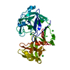


 PDBj
PDBj


 Assembly
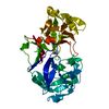

 Mass: 46.068 Da / Num. of mol.: 2 / Source method: obtained synthetically / Formula: C2H6O
 Mass: 18.015 Da / Num. of mol.: 206 / Source method: isolated from a natural source / Formula: H2O
 Sample preparation
 Processing