 Movie
Movie Controller


+ Open data

- Basic information
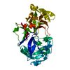
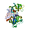
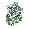
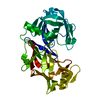
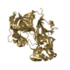
| Entry | Database: PDB / ID: 2jxr | ||||||||||||
|---|---|---|---|---|---|---|---|---|---|---|---|---|---|
| Title | STRUCTURE OF YEAST PROTEINASE A | ||||||||||||
 Components Components | PROTEINASE A | ||||||||||||
 Keywords Keywords | HYDROLASE/HYDROLASE INHIBITOR / HYDROLASE-HYDROLASE INHIBITOR COMPLEX / ASPARTYL PROTEASE / GLYCOPROTEIN / ZYMOGEN | ||||||||||||
| Function / homology |  Function and homology information Function and homology informationsaccharopepsin / microautophagy / cytoplasm to vacuole targeting by the Cvt pathway / oligosaccharide binding / fungal-type vacuole / pexophagy / : / macroautophagy / autophagy / disordered domain specific binding ...saccharopepsin / microautophagy / cytoplasm to vacuole targeting by the Cvt pathway / oligosaccharide binding / fungal-type vacuole / pexophagy / : / macroautophagy / autophagy / disordered domain specific binding / peptidase activity / aspartic-type endopeptidase activity / endoplasmic reticulum / protein-containing complex / mitochondrion Similarity search - Function | ||||||||||||
| Biological species |  | ||||||||||||
| Method |  X-RAY DIFFRACTION / SYNCHROTRON / Resolution: 2.4 Å X-RAY DIFFRACTION / SYNCHROTRON / Resolution: 2.4 Å | ||||||||||||
 Authors Authors | Aguilar, C.F. / Badasso, M. / Dreyer, T. / Cronin, N.B. / Newman, M.P. / Cooper, J.B. / Hoover, D.J. / Wood, S.P. / Johnson, M.S. / Blundell, T.L. | ||||||||||||
 Citation Citation | Journal: J.Mol.Biol. / Year: 1997 Title: The three-dimensional structure at 2.4 A resolution of glycosylated proteinase A from the lysosome-like vacuole of Saccharomyces cerevisiae. Authors: Aguilar, C.F. / Cronin, N.B. / Badasso, M. / Dreyer, T. / Newman, M.P. / Cooper, J.B. / Hoover, D.J. / Wood, S.P. / Johnson, M.S. / Blundell, T.L. | ||||||||||||
| History |
|
- Structure visualization
| Structure viewer | Molecule: MolmilJmol/JSmol |
|---|
- Downloads & links
-Download
| PDBx/mmCIF format | 2jxr.cif.gz | 86.3 KB | Display | PDBx/mmCIF format |
|---|---|---|---|---|
| PDB format | pdb2jxr.ent.gz | 62.5 KB | Display | PDB format |
| PDBx/mmJSON format | 2jxr.json.gz | Tree view | PDBx/mmJSON format | |
| Others |  Other downloads Other downloads |
-Validation report
| Arichive directory | https://data.pdbj.org/pub/pdb/validation_reports/jx/2jxrftp://data.pdbj.org/pub/pdb/validation_reports/jx/2jxr | HTTPS FTP |
|---|
-Related structure data
| Similar structure data |
|---|


-Links
 PDBj
PDBj- Assembly
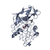
| Deposited unit | 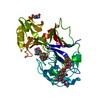
| ||||||||
|---|---|---|---|---|---|---|---|---|---|
| 1 |
| ||||||||
| 2 | 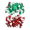
| ||||||||
| Unit cell |
|
-Components
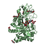
| #1: Protein | Mass: 35774.551 Da / Num. of mol.: 1 / Mutation: L315I Source method: isolated from a genetically manipulated source Source: (gene. exp.) References: UniProt: P07267, saccharopepsin |
|---|---|
| #2: Polysaccharide | alpha-D-mannopyranose-(1-2)-beta-D-mannopyranose-(1-3)-beta-D-mannopyranose-(1-4)-2-acetamido-2- ...alpha-D-mannopyranose-(1-2)-beta-D-mannopyranose-(1-3)-beta-D-mannopyranose-(1-4)-2-acetamido-2-deoxy-beta-D-glucopyranose-(1-4)-2-acetamido-2-deoxy-beta-D-glucopyranose Source method: isolated from a genetically manipulated source |
| #3: Chemical | ChemComp-2Z3 / 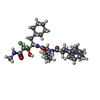  Type: peptide-like, Peptide-like / Class: Inhibitor / Mass: 653.758 Da / Num. of mol.: 1 / Source method: obtained synthetically / Formula: C32H49F2N5O7 / References: CP-81,282, MOR-PHE-NLE-CHF-NME Type: peptide-like, Peptide-like / Class: Inhibitor / Mass: 653.758 Da / Num. of mol.: 1 / Source method: obtained synthetically / Formula: C32H49F2N5O7 / References: CP-81,282, MOR-PHE-NLE-CHF-NME |
| #4: Sugar | ChemComp-NAG /   Type: D-saccharide, beta linking / Mass: 221.208 Da / Num. of mol.: 1 Type: D-saccharide, beta linking / Mass: 221.208 Da / Num. of mol.: 1Source method: isolated from a genetically manipulated source Formula: C8H15NO6 |
| #5: Water | ChemComp-HOH /  Mass: 18.015 Da / Num. of mol.: 119 / Source method: isolated from a natural source / Formula: H2O Mass: 18.015 Da / Num. of mol.: 119 / Source method: isolated from a natural source / Formula: H2O |
| Has protein modification | Y |
-Experimental details
-Experiment
| Experiment | Method: X-RAY DIFFRACTION |
|---|
- Sample preparation
| Crystal | Density Matthews: 3.3 Å3/Da / Density % sol: 63 % | ||||||||||||||||||||
|---|---|---|---|---|---|---|---|---|---|---|---|---|---|---|---|---|---|---|---|---|---|
| Crystal grow | *PLUS Method: vapor diffusion / Details: Badasso, M., (1993) J. Mol. Biol., 232, 701. | ||||||||||||||||||||
| Components of the solutions | *PLUS
|
-Data collection
| Diffraction source | Source: SYNCHROTRON / Site: Photon Factory  / Beamline: BL-6A / Wavelength: 1 / Beamline: BL-6A / Wavelength: 1 |
|---|---|
| Detector | Detector: IMAGE PLATE / Date: Nov 1, 1993 |
| Radiation | Monochromatic (M) / Laue (L): M / Scattering type: x-ray |
| Radiation wavelength | Wavelength: 1 Å / Relative weight: 1 |
| Reflection | Num. obs: 18325 / % possible obs: 95 % / Rmerge(I) obs: 0.07 |
| Reflection | *PLUS Highest resolution: 2.4 Å / Num. measured all: 84192 |
- Processing
| Software |
| ||||||||||||||||||||||||
|---|---|---|---|---|---|---|---|---|---|---|---|---|---|---|---|---|---|---|---|---|---|---|---|---|---|
| Refinement | Resolution: 2.4→10 Å Details: LOOP RESIDUES A 140 - A 142 WERE MODELED STEREOCHEMICALLY.
| ||||||||||||||||||||||||
| Displacement parameters | Biso mean: 37.01 Å2 | ||||||||||||||||||||||||
| Refinement step | Cycle: LAST / Resolution: 2.4→10 Å
| ||||||||||||||||||||||||
| Software | *PLUS Name: RESTRAIN / Classification: refinement | ||||||||||||||||||||||||
| Refinement | *PLUS Rfactor all: 0.193 / Rfactor Rfree: 0.27 | ||||||||||||||||||||||||
| Solvent computation | *PLUS | ||||||||||||||||||||||||
| Displacement parameters | *PLUS | ||||||||||||||||||||||||
| Refine LS restraints | *PLUS
|
