 Movie
Movie Controller


+ Open data

- Basic information
| Entry | Database: PDB / ID: 3tlk | ||||||
|---|---|---|---|---|---|---|---|
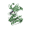
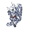
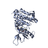
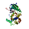
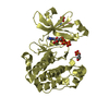
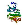
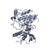
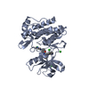
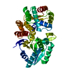
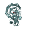
| Title | Crystal structure of holo FepB | ||||||
 Components Components | Ferrienterobactin-binding periplasmic protein | ||||||
 Keywords Keywords | METAL TRANSPORT / Ferric-enterobactin / trimer / siderophore transport / Periplasmic space | ||||||
| Function / homology |  Function and homology information Function and homology informationferric-enterobactin import into cell / iron import into cell / Iron assimilation using enterobactin / outer membrane-bounded periplasmic space / DNA damage response / membrane Similarity search - Function | ||||||
| Biological species |  | ||||||
| Method |  X-RAY DIFFRACTION / SYNCHROTRON / MOLECULAR REPLACEMENT / Resolution: 1.853 Å X-RAY DIFFRACTION / SYNCHROTRON / MOLECULAR REPLACEMENT / Resolution: 1.853 Å | ||||||
 Authors Authors | Li, N. / Gu, L. | ||||||
 Citation Citation | Journal: To be Published Title: Crystal structure of holo FepB Authors: Li, N. / Gu, L. | ||||||
| History |
|
- Structure visualization
| Structure viewer | Molecule: MolmilJmol/JSmol |
|---|
- Downloads & links
-Download
| PDBx/mmCIF format | 3tlk.cif.gz | 199.1 KB | Display | PDBx/mmCIF format |
|---|---|---|---|---|
| PDB format | pdb3tlk.ent.gz | 156.3 KB | Display | PDB format |
| PDBx/mmJSON format | 3tlk.json.gz | Tree view | PDBx/mmJSON format | |
| Others |  Other downloads Other downloads |
-Validation report
| Arichive directory | https://data.pdbj.org/pub/pdb/validation_reports/tl/3tlkftp://data.pdbj.org/pub/pdb/validation_reports/tl/3tlk | HTTPS FTP |
|---|
-Related structure data
| Related structure data |  3r5tS S: Starting model for refinement |
|---|---|
| Similar structure data |
-Links
 PDBj
PDBj
- Assembly
| Deposited unit | 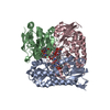
| ||||||||||||
|---|---|---|---|---|---|---|---|---|---|---|---|---|---|
| 1 | 
| ||||||||||||
| 2 | 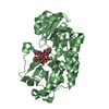
| ||||||||||||
| 3 | 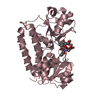
| ||||||||||||
| Unit cell |
| ||||||||||||
| Components on special symmetry positions |
|
-Components
| #1: Protein | Mass: 35385.043 Da / Num. of mol.: 3 / Fragment: periplasmic transport domain Source method: isolated from a genetically manipulated source Source: (gene. exp.) #2: Chemical | ChemComp-FE /   Mass: 55.845 Da / Num. of mol.: 4 / Source method: obtained synthetically / Formula: Fe Mass: 55.845 Da / Num. of mol.: 4 / Source method: obtained synthetically / Formula: Fe#3: Chemical | ChemComp-EB4 /   Mass: 669.546 Da / Num. of mol.: 4 / Source method: obtained synthetically / Formula: C30H27N3O15 Mass: 669.546 Da / Num. of mol.: 4 / Source method: obtained synthetically / Formula: C30H27N3O15#4: Chemical |   Mass: 88.105 Da / Num. of mol.: 3 / Source method: obtained synthetically / Formula: C4H8O2 Mass: 88.105 Da / Num. of mol.: 3 / Source method: obtained synthetically / Formula: C4H8O2#5: Water | ChemComp-HOH / |  Mass: 18.015 Da / Num. of mol.: 616 / Source method: isolated from a natural source / Formula: H2O Mass: 18.015 Da / Num. of mol.: 616 / Source method: isolated from a natural source / Formula: H2O |
|---|
-Experimental details
-Experiment
| Experiment | Method: X-RAY DIFFRACTION / Number of used crystals: 1 |
|---|
- Sample preparation
| Crystal | Density Matthews: 2.25 Å3/Da / Density % sol: 45.32 % |
|---|---|
| Crystal grow | Temperature: 293 K / Method: vapor diffusion, sitting drop / pH: 7 Details: 1.0M Succinic acid pH 7.0, 0.1M HEPES, 1% w/v Polyethylene glycol monomethyl ether 2000, VAPOR DIFFUSION, SITTING DROP, temperature 293K |
-Data collection
| Diffraction | Mean temperature: 100 K | |||||||||||||||||||||
|---|---|---|---|---|---|---|---|---|---|---|---|---|---|---|---|---|---|---|---|---|---|---|
| Diffraction source | Source: SYNCHROTRON / Site: SSRF  / Beamline: BL17U / Wavelength: 0.9792 Å / Beamline: BL17U / Wavelength: 0.9792 Å | |||||||||||||||||||||
| Detector | Type: MARMOSAIC 225 mm CCD / Detector: CCD / Date: Jan 31, 2010 | |||||||||||||||||||||
| Radiation | Monochromator: SAGITALLY FOCUSED Si(111) / Protocol: SINGLE WAVELENGTH / Monochromatic (M) / Laue (L): M / Scattering type: x-ray | |||||||||||||||||||||
| Radiation wavelength | Wavelength: 0.9792 Å / Relative weight: 1 | |||||||||||||||||||||
| Reflection | Resolution: 1.85→50 Å / Num. all: 79154 / Num. obs: 79154 / % possible obs: 99.9 % / Observed criterion σ(F): 0 / Observed criterion σ(I): 0 / Redundancy: 6.7 % / Rmerge(I) obs: 0.104 / Rsym value: 0.104 / Net I/σ(I): 19.17 | |||||||||||||||||||||
| Reflection shell | Diffraction-ID: 1
|
- Processing
| Software |
| ||||||||||||||||||||||||||||||||||||||||||||||||||||||||||||||||||||||||||||||||||||
|---|---|---|---|---|---|---|---|---|---|---|---|---|---|---|---|---|---|---|---|---|---|---|---|---|---|---|---|---|---|---|---|---|---|---|---|---|---|---|---|---|---|---|---|---|---|---|---|---|---|---|---|---|---|---|---|---|---|---|---|---|---|---|---|---|---|---|---|---|---|---|---|---|---|---|---|---|---|---|---|---|---|---|---|---|---|
| Refinement | Method to determine structure: MOLECULAR REPLACEMENT Starting model: 3R5T Resolution: 1.853→39.314 Å / SU ML: 0.21 / σ(F): 0.04 / Phase error: 21.61 / Stereochemistry target values: ML
| ||||||||||||||||||||||||||||||||||||||||||||||||||||||||||||||||||||||||||||||||||||
| Solvent computation | Shrinkage radii: 0.9 Å / VDW probe radii: 1.11 Å / Solvent model: FLAT BULK SOLVENT MODEL / Bsol: 40.687 Å2 / ksol: 0.378 e/Å3 | ||||||||||||||||||||||||||||||||||||||||||||||||||||||||||||||||||||||||||||||||||||
| Displacement parameters | Biso mean: 28.108 Å2
| ||||||||||||||||||||||||||||||||||||||||||||||||||||||||||||||||||||||||||||||||||||
| Refinement step | Cycle: LAST / Resolution: 1.853→39.314 Å
| ||||||||||||||||||||||||||||||||||||||||||||||||||||||||||||||||||||||||||||||||||||
| Refine LS restraints |
| ||||||||||||||||||||||||||||||||||||||||||||||||||||||||||||||||||||||||||||||||||||
| LS refinement shell | Refine-ID: X-RAY DIFFRACTION / Total num. of bins used: 13
|
