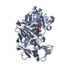
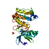
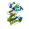
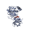
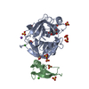
Entry Database : PDB / ID : 3oz0Title PPAR Delta in complex with azppard02 Peroxisome proliferator-activated receptor delta Keywords / / / / / Function / homology Function Domain/homology Component
/ / / / / / / / / / / / / / / / / / / / / / / / / / / / / / / / / / / / / / / / / / / / / / / / / / / / / / / / / / / / / / / / / / / / / / / / / / / / / / / / / / / / / / / / / / / / / / / / / / / / / / / / / / / / / / / / / / / / / Biological species Homo sapiens (human)Method / / OTHER / Resolution : 3 Å Authors Ogg, D. Journal : Bioorg.Med.Chem.Lett. / Year : 2011Title : Discovery of isoindoline and tetrahydroisoquinoline derivatives as potent, selective PPARδ agonistsAuthors : Luckhurst, C.A. / Stein, L.A. / Furber, M. / Webb, N. / Ratcliffe, M.J. / Allenby, G. / Botterell, S. / Tomlinson, W. / Martin, B. / Walding, A. History Deposition Sep 24, 2010 Deposition site / Processing site Revision 1.0 Jan 19, 2011 Provider / Type Revision 1.1 Jul 13, 2011 Group Revision 1.2 Feb 29, 2012 Group Revision 1.3 Nov 8, 2017 Group / Refinement description / Category / software / Item Revision 1.4 Sep 6, 2023 Group Advisory / Data collection ... Advisory / Data collection / Database references / Derived calculations / Refinement description Category chem_comp_atom / chem_comp_bond ... chem_comp_atom / chem_comp_bond / database_2 / pdbx_initial_refinement_model / pdbx_unobs_or_zero_occ_atoms / struct_ref_seq_dif / struct_site Item _database_2.pdbx_DOI / _database_2.pdbx_database_accession ... _database_2.pdbx_DOI / _database_2.pdbx_database_accession / _struct_ref_seq_dif.details / _struct_site.pdbx_auth_asym_id / _struct_site.pdbx_auth_comp_id / _struct_site.pdbx_auth_seq_id
Show all Show less
 Movie
Movie Controller


 Open data
 Basic information
 Components
 Keywords
 Function and homology information
 Homo sapiens (human)
 X-RAY DIFFRACTION /
  Authors
 Citation
 Structure visualization
 Downloads & links
 Other downloads


 PDBj
PDBj
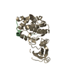
 Assembly


 Mass: 485.359 Da / Num. of mol.: 1 / Source method: obtained synthetically / Formula: C25H22Cl2N2O4
 Sample preparation
 / Beamline: X06SA / Wavelength: 0.9134 / Wavelength: 0.9134 Å
 Processing