 Movie
Movie Controller

[English] 日本語
 Yorodumi
- PDB-1al8: THREE-DIMENSIONAL STRUCTURE OF GLYCOLATE OXIDASE WITH BOUND ACTIV... -
+ Open data

- Basic information
| Entry | Database: PDB / ID: 1al8 | ||||||
|---|---|---|---|---|---|---|---|
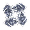
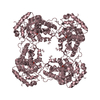
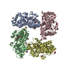
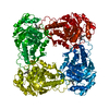
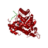
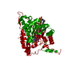
| Title | THREE-DIMENSIONAL STRUCTURE OF GLYCOLATE OXIDASE WITH BOUND ACTIVE-SITE INHIBITORS | ||||||
 Components Components | GLYCOLATE OXIDASE | ||||||
 Keywords Keywords | FLAVOPROTEIN / DRUG DESIGN / INHIBITOR BINDING | ||||||
| Function / homology |  Function and homology information Function and homology informationoxidative photosynthetic carbon pathway / (S)-2-hydroxy-acid oxidase / (S)-2-hydroxy-acid oxidase activity / hydrogen peroxide biosynthetic process / FMN binding / peroxisome Similarity search - Function | ||||||
| Biological species |  Spinacia oleracea (spinach) Spinacia oleracea (spinach) | ||||||
| Method |  X-RAY DIFFRACTION / MOLECULAR REPLACEMENT / Resolution: 2.2 Å X-RAY DIFFRACTION / MOLECULAR REPLACEMENT / Resolution: 2.2 Å | ||||||
 Authors Authors | Stenberg, K. / Lindqvist, Y. | ||||||
 Citation Citation | Journal: Protein Sci. / Year: 1997 Title: Three-dimensional structures of glycolate oxidase with bound active-site inhibitors. Authors: Stenberg, K. / Lindqvist, Y. #1: Journal: Protein Expr.Purif. / Year: 1996Title: High-Level Expression, Purification and Crystallization of Recombinant Spinach Glycolate Oxidase in Escherichia Coli Authors: Stenberg, K. / Lindqvist, Y. #2: Journal: J.Mol.Biol. / Year: 1989Title: Refined Structure of Spinach Glycolate Oxidase at 2 A Resolution Authors: Lindqvist, Y. | ||||||
| History |
|
- Structure visualization
| Structure viewer | Molecule: MolmilJmol/JSmol |
|---|
- Downloads & links
-Download
| PDBx/mmCIF format | 1al8.cif.gz | 81.7 KB | Display | PDBx/mmCIF format |
|---|---|---|---|---|
| PDB format | pdb1al8.ent.gz | 61.3 KB | Display | PDB format |
| PDBx/mmJSON format | 1al8.json.gz | Tree view | PDBx/mmJSON format | |
| Others |  Other downloads Other downloads |
-Validation report
| Arichive directory | https://data.pdbj.org/pub/pdb/validation_reports/al/1al8ftp://data.pdbj.org/pub/pdb/validation_reports/al/1al8 | HTTPS FTP |
|---|
-Related structure data
| Related structure data |  1al7C 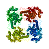 1goxS S: Starting model for refinement C: citing same article ( |
|---|---|
| Similar structure data |


-Links
 PDBj
PDBj
- Assembly
| Deposited unit | 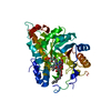
| ||||||||
|---|---|---|---|---|---|---|---|---|---|
| 1 | 
| ||||||||
| Unit cell |
|
-Components
| #1: Protein | Mass: 39336.352 Da / Num. of mol.: 1 Source method: isolated from a genetically manipulated source Details: THE STRUCTURE DESCRIBED IS A COMPLEX OF GLYCOLATE OXIDASE WITH FMN AND AN INHIBITOR Source: (gene. exp.) Spinacia oleracea (spinach) / Cell line: BL21 / Cellular location: PEROXISOME / Plasmid: PKS20+ / Cellular location (production host): CYTOPLASM / Production host:  |
|---|---|
| #2: Chemical | ChemComp-FMN /   Mass: 456.344 Da / Num. of mol.: 1 / Source method: obtained synthetically / Formula: C17H21N4O9P Mass: 456.344 Da / Num. of mol.: 1 / Source method: obtained synthetically / Formula: C17H21N4O9P |
| #3: Chemical | ChemComp-DHP /   Type: L-peptide linking / Mass: 253.337 Da / Num. of mol.: 1 / Source method: obtained synthetically / Formula: C14H23NO3 Type: L-peptide linking / Mass: 253.337 Da / Num. of mol.: 1 / Source method: obtained synthetically / Formula: C14H23NO3 |
| #4: Water | ChemComp-HOH /  Mass: 18.015 Da / Num. of mol.: 68 / Source method: isolated from a natural source / Formula: H2O Mass: 18.015 Da / Num. of mol.: 68 / Source method: isolated from a natural source / Formula: H2O |
-Experimental details
-Experiment
| Experiment | Method: X-RAY DIFFRACTION / Number of used crystals: 1 |
|---|
- Sample preparation
| Crystal | Density Matthews: 2.5 Å3/Da / Density % sol: 54.5 % | ||||||||||||||||||||||||||||||||||||||||
|---|---|---|---|---|---|---|---|---|---|---|---|---|---|---|---|---|---|---|---|---|---|---|---|---|---|---|---|---|---|---|---|---|---|---|---|---|---|---|---|---|---|
| Crystal grow | pH: 8.3 Details: PROTEIN WAS CRYSTALLIZED FROM 50MM TRIS- BUFFER, 0.25 MG/ML FMN, 4% TERTIARY BUTANOL AND 0.5 % JF 5969, PH 8.3. JF 5969 IS A MIXTURE OF SYMPERONIC NPE1800 (33.3G/L), TWEEN 85 (20G/L) IN CYCLOHEXANONE. | ||||||||||||||||||||||||||||||||||||||||
| Crystal grow | *PLUS Method: microdialysis | ||||||||||||||||||||||||||||||||||||||||
| Components of the solutions | *PLUS
|
-Data collection
| Diffraction | Mean temperature: 277 K |
|---|---|
| Diffraction source | Wavelength: 1.5418 |
| Detector | Type: MARRESEARCH / Detector: IMAGE PLATE / Date: Apr 1, 1996 |
| Radiation | Monochromatic (M) / Laue (L): M / Scattering type: x-ray |
| Radiation wavelength | Wavelength: 1.5418 Å / Relative weight: 1 |
| Reflection | Resolution: 2.2→100 Å / Num. obs: 21308 / % possible obs: 99.8 % / Observed criterion σ(I): 0 / Redundancy: 11.1 % / Biso Wilson estimate: 0.317 Å2 / Rsym value: 0.094 / Net I/σ(I): 7.9 |
| Reflection shell | Resolution: 2.2→2.25 Å / Mean I/σ(I) obs: 3.3 / Rsym value: 0.369 / % possible all: 99.9 |
| Reflection | *PLUS Num. measured all: 237398 / Rmerge(I) obs: 0.094 |
| Reflection shell | *PLUS % possible obs: 99.9 % / Rmerge(I) obs: 0.369 |
- Processing
| Software |
| ||||||||||||||||||||||||||||||||||||||||||||||||||||||||||||
|---|---|---|---|---|---|---|---|---|---|---|---|---|---|---|---|---|---|---|---|---|---|---|---|---|---|---|---|---|---|---|---|---|---|---|---|---|---|---|---|---|---|---|---|---|---|---|---|---|---|---|---|---|---|---|---|---|---|---|---|---|---|
| Refinement | Method to determine structure: MOLECULAR REPLACEMENT Starting model: PDB ENTRY 1GOX Resolution: 2.2→8 Å / Data cutoff low absF: 0 / Isotropic thermal model: INDIVIDUAL / Cross valid method: THROUGHOUT / σ(F): 0 Details: TOPOLOGY AND PARAMETER FILES FOR THE TKP-INHIBITOR WERE CREATED USING THE PROGRAM XPLO2D (REF: [O/X-PLOR DICTIONARIES] G.J. KLEYWEGT, DICTIONARIES FOR HETEROS, ESF/CCP4 NEWSLETTER 31,JUNE ...Details: TOPOLOGY AND PARAMETER FILES FOR THE TKP-INHIBITOR WERE CREATED USING THE PROGRAM XPLO2D (REF: [O/X-PLOR DICTIONARIES] G.J. KLEYWEGT, DICTIONARIES FOR HETEROS, ESF/CCP4 NEWSLETTER 31,JUNE 1995, PP. 45-50). BOND LENGTHS AND ANGLES ARE FROM IDEALIZED STRUCTURES.
| ||||||||||||||||||||||||||||||||||||||||||||||||||||||||||||
| Displacement parameters | Biso mean: 0.276 Å2 | ||||||||||||||||||||||||||||||||||||||||||||||||||||||||||||
| Refinement step | Cycle: LAST / Resolution: 2.2→8 Å
| ||||||||||||||||||||||||||||||||||||||||||||||||||||||||||||
| Refine LS restraints |
| ||||||||||||||||||||||||||||||||||||||||||||||||||||||||||||
| LS refinement shell | Resolution: 2.2→2.34 Å / Total num. of bins used: 12 /
| ||||||||||||||||||||||||||||||||||||||||||||||||||||||||||||
| Xplor file |
| ||||||||||||||||||||||||||||||||||||||||||||||||||||||||||||
| Software | *PLUS Name: X-PLOR / Version: 3.1F / Classification: refinement | ||||||||||||||||||||||||||||||||||||||||||||||||||||||||||||
| Refinement | *PLUS | ||||||||||||||||||||||||||||||||||||||||||||||||||||||||||||
| Solvent computation | *PLUS | ||||||||||||||||||||||||||||||||||||||||||||||||||||||||||||
| Displacement parameters | *PLUS Biso mean: 27.6 Å2 | ||||||||||||||||||||||||||||||||||||||||||||||||||||||||||||
| Refine LS restraints | *PLUS
| ||||||||||||||||||||||||||||||||||||||||||||||||||||||||||||
| LS refinement shell | *PLUS Rfactor obs: 0.265 |
