 Movie
Movie Controller

[English] 日本語
 Yorodumi
- PDB-1ac6: CRYSTAL STRUCTURE OF A VARIABLE DOMAIN MUTANT OF A T-CELL RECEPTO... -
+ Open data

- Basic information
| Entry | Database: PDB / ID: 1ac6 | ||||||
|---|---|---|---|---|---|---|---|
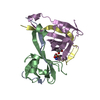
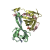
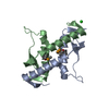
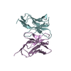
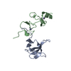
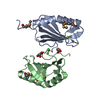
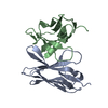
| Title | CRYSTAL STRUCTURE OF A VARIABLE DOMAIN MUTANT OF A T-CELL RECEPTOR ALPHA CHAIN | ||||||
 Components Components | T-CELL RECEPTOR ALPHA | ||||||
 Keywords Keywords | RECEPTOR / V ALPHA DOMAIN / SITE-DIRECTED MUTAGENESIS / THREE-DIMENSIONAL STRUCTURE / GLYCOPROTEIN | ||||||
| Function / homology |  Function and homology information Function and homology information | ||||||
| Biological species |  | ||||||
| Method |  X-RAY DIFFRACTION / MOLECULAR REPLACEMENT / Resolution: 2.3 Å X-RAY DIFFRACTION / MOLECULAR REPLACEMENT / Resolution: 2.3 Å | ||||||
 Authors Authors | Li, H.-M. / Mariuzza, R.A. | ||||||
 Citation Citation | Journal: J.Mol.Biol. / Year: 1997 Title: Dual conformations of a T cell receptor V alpha homodimer: implications for variability in V alpha V beta domain association. Authors: Li, H. / Lebedeva, M.I. / Ward, E.S. / Mariuzza, R.A. #1: Journal: Nature / Year: 1996Title: Crystal Structure of a T-Cell Receptor Beta-Chain Complexed with a Superantigen Authors: Fields, B.A. / Malchiodi, E.L. / Li, H. / Ysern, X. / Stauffacher, C.V. / Schlievert, P.M. / Karjalainen, K. / Mariuzza, R.A. #2: Journal: Science / Year: 1995Title: Crystal Structure of the Beta Chain of a T Cell Antigen Receptor Authors: Bentley, G.A. / Boulot, G. / Karjalainen, K. / Mariuzza, R.A. #3: Journal: Science / Year: 1995Title: Crystal Structure of the V Alpha Domain of a T Cell Antigen Receptor Authors: Fields, B.A. / Ober, B. / Malchiodi, E.L. / Lebedeva, M.I. / Braden, B.C. / Ysern, X. / Kim, J.K. / Shao, X. / Ward, E.S. / Mariuzza, R.A. | ||||||
| History |
|
- Structure visualization
| Structure viewer | Molecule: MolmilJmol/JSmol |
|---|
- Downloads & links
-Download
| PDBx/mmCIF format | 1ac6.cif.gz | 57.1 KB | Display | PDBx/mmCIF format |
|---|---|---|---|---|
| PDB format | pdb1ac6.ent.gz | 41.5 KB | Display | PDB format |
| PDBx/mmJSON format | 1ac6.json.gz | Tree view | PDBx/mmJSON format | |
| Others |  Other downloads Other downloads |
-Validation report
| Arichive directory | https://data.pdbj.org/pub/pdb/validation_reports/ac/1ac6ftp://data.pdbj.org/pub/pdb/validation_reports/ac/1ac6 | HTTPS FTP |
|---|
-Related structure data
| Similar structure data |
|---|


-Links
 PDBj
PDBj- Assembly
| Deposited unit | 
| ||||||||
|---|---|---|---|---|---|---|---|---|---|
| 1 |
| ||||||||
| Unit cell |
| ||||||||
| Noncrystallographic symmetry (NCS) | NCS oper: (Code: given Matrix: (-0.86457, -0.497718, 0.069259), Vector: |
-Components
| #1: Protein | Mass: 12104.274 Da / Num. of mol.: 2 / Fragment: VARIABLE DOMAIN / Mutation: yes / Source method: isolated from a natural source / Source: (natural) #2: Water | ChemComp-HOH / |  Mass: 18.015 Da / Num. of mol.: 116 / Source method: isolated from a natural source / Formula: H2O Mass: 18.015 Da / Num. of mol.: 116 / Source method: isolated from a natural source / Formula: H2OHas protein modification | Y | |
|---|
-Experimental details
-Experiment
| Experiment | Method: X-RAY DIFFRACTION / Number of used crystals: 1 |
|---|
- Sample preparation
| Crystal | Density Matthews: 2.1 Å3/Da / Density % sol: 42 % | ||||||||||||||||||||
|---|---|---|---|---|---|---|---|---|---|---|---|---|---|---|---|---|---|---|---|---|---|
| Crystal grow | Method: vapor diffusion, hanging drop / pH: 7.7 Details: PROTEIN WAS CRYSTALLIZED FROM 5.0M SODIUM FORMATE, PH 7.7 IN HANGING DROPS AT ROOM TEMPERATURE., vapor diffusion - hanging drop Temp details: room temp | ||||||||||||||||||||
| Crystal grow | *PLUS Method: vapor diffusion, hanging drop | ||||||||||||||||||||
| Components of the solutions | *PLUS
|
-Data collection
| Diffraction | Mean temperature: 293 K |
|---|---|
| Diffraction source | Source: ROTATING ANODE / Type: RIGAKU RUH2R / Wavelength: 1.5418 |
| Detector | Type: SIEMENS / Detector: AREA DETECTOR / Date: Nov 1, 1995 / Details: COLLIMATOR |
| Radiation | Monochromator: NI FILTER / Monochromatic (M) / Laue (L): M / Scattering type: x-ray |
| Radiation wavelength | Wavelength: 1.5418 Å / Relative weight: 1 |
| Reflection | Resolution: 2.12→26.41 Å / Num. obs: 9957 / % possible obs: 78.2 % / Observed criterion σ(I): -3 / Redundancy: 3 % / Rmerge(I) obs: 0.105 / Rsym value: 0.105 / Net I/σ(I): 6 |
| Reflection shell | Resolution: 2.3→2.37 Å / Redundancy: 2.7 % / Rmerge(I) obs: 0.406 / Mean I/σ(I) obs: 1.38 / Rsym value: 0.406 / % possible all: 73 |
| Reflection | *PLUS Num. measured all: 26767 |
| Reflection shell | *PLUS % possible obs: 72.7 % |
- Processing
| Software |
| ||||||||||||||||||||||||||||||||||||||||||||||||||||||||||||
|---|---|---|---|---|---|---|---|---|---|---|---|---|---|---|---|---|---|---|---|---|---|---|---|---|---|---|---|---|---|---|---|---|---|---|---|---|---|---|---|---|---|---|---|---|---|---|---|---|---|---|---|---|---|---|---|---|---|---|---|---|---|
| Refinement | Method to determine structure: MOLECULAR REPLACEMENT Starting model: VALPHA WILDTYPE HOMODIMER STRUCTURE OF 1934.4 TCR Resolution: 2.3→8 Å / Data cutoff high absF: 10000000 / Data cutoff low absF: 0 / Isotropic thermal model: ISOTROPIC / Cross valid method: FREE R / σ(F): 2
| ||||||||||||||||||||||||||||||||||||||||||||||||||||||||||||
| Displacement parameters | Biso mean: 24.1 Å2 | ||||||||||||||||||||||||||||||||||||||||||||||||||||||||||||
| Refine analyze | Luzzati coordinate error obs: 0.23 Å | ||||||||||||||||||||||||||||||||||||||||||||||||||||||||||||
| Refinement step | Cycle: LAST / Resolution: 2.3→8 Å
| ||||||||||||||||||||||||||||||||||||||||||||||||||||||||||||
| Refine LS restraints |
| ||||||||||||||||||||||||||||||||||||||||||||||||||||||||||||
| LS refinement shell | Resolution: 2.3→2.35 Å / Total num. of bins used: 15
| ||||||||||||||||||||||||||||||||||||||||||||||||||||||||||||
| Xplor file |
| ||||||||||||||||||||||||||||||||||||||||||||||||||||||||||||
| Software | *PLUS Name: X-PLOR / Version: 3.1 / Classification: refinement | ||||||||||||||||||||||||||||||||||||||||||||||||||||||||||||
| Refinement | *PLUS Num. reflection all: 7850 / Rfactor all: 0.198 | ||||||||||||||||||||||||||||||||||||||||||||||||||||||||||||
| Solvent computation | *PLUS | ||||||||||||||||||||||||||||||||||||||||||||||||||||||||||||
| Displacement parameters | *PLUS | ||||||||||||||||||||||||||||||||||||||||||||||||||||||||||||
| Refine LS restraints | *PLUS
|
