 Movie
Movie Controller

[English] 日本語
 Yorodumi
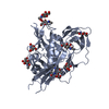
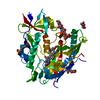
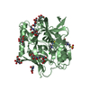
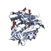
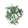
- PDB-1a9n: CRYSTAL STRUCTURE OF THE SPLICEOSOMAL U2B''-U2A' PROTEIN COMPLEX ... -
+ Open data

- Basic information
| Entry | Database: PDB / ID: 1a9n | ||||||
|---|---|---|---|---|---|---|---|
| Title | CRYSTAL STRUCTURE OF THE SPLICEOSOMAL U2B''-U2A' PROTEIN COMPLEX BOUND TO A FRAGMENT OF U2 SMALL NUCLEAR RNA | ||||||
 Components Components |
| ||||||
 Keywords Keywords | RNA BINDING PROTEIN/RNA / COMPLEX (NUCLEAR PROTEIN-RNA) / RNA / SNRNP / RIBONUCLEOPROTEIN / RNA BINDING PROTEIN-RNA COMPLEX | ||||||
| Function / homology |  Function and homology information Function and homology informationsmall nuclear ribonucleoprotein complex / U2-type precatalytic spliceosome / U2-type prespliceosome assembly / U2-type catalytic step 2 spliceosome / U2-type spliceosomal complex / U1 snRNP / U2 snRNP / U2 snRNA binding / U1 snRNA binding / catalytic step 2 spliceosome ...small nuclear ribonucleoprotein complex / U2-type precatalytic spliceosome / U2-type prespliceosome assembly / U2-type catalytic step 2 spliceosome / U2-type spliceosomal complex / U1 snRNP / U2 snRNP / U2 snRNA binding / U1 snRNA binding / catalytic step 2 spliceosome / mRNA Splicing - Major Pathway / Gene and protein expression by JAK-STAT signaling after Interleukin-12 stimulation / RNA splicing / spliceosomal complex / mRNA splicing, via spliceosome / fibrillar center / cytoplasmic ribonucleoprotein granule / spermatogenesis / nuclear speck / nuclear body / RNA binding / nucleoplasm / nucleus Similarity search - Function | ||||||
| Biological species |  Homo sapiens (human) Homo sapiens (human) | ||||||
| Method |  X-RAY DIFFRACTION / SYNCHROTRON / SIR / Resolution: 2.38 Å X-RAY DIFFRACTION / SYNCHROTRON / SIR / Resolution: 2.38 Å | ||||||
 Authors Authors | Price, S.R. / Evans, P.R. / Nagai, K. | ||||||
 Citation Citation | Journal: Nature / Year: 1998 Title: Crystal structure of the spliceosomal U2B"-U2A' protein complex bound to a fragment of U2 small nuclear RNA. Authors: Price, S.R. / Evans, P.R. / Nagai, K. | ||||||
| History |
|
- Structure visualization
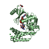
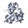
| Structure viewer | Molecule: MolmilJmol/JSmol |
|---|
- Downloads & links
-Download
| PDBx/mmCIF format | 1a9n.cif.gz | 140.5 KB | Display | PDBx/mmCIF format |
|---|---|---|---|---|
| PDB format | pdb1a9n.ent.gz | 108.7 KB | Display | PDB format |
| PDBx/mmJSON format | 1a9n.json.gz | Tree view | PDBx/mmJSON format | |
| Others |  Other downloads Other downloads |
-Validation report
| Arichive directory | https://data.pdbj.org/pub/pdb/validation_reports/a9/1a9nftp://data.pdbj.org/pub/pdb/validation_reports/a9/1a9n | HTTPS FTP |
|---|
-Related structure data
| Similar structure data |
|---|


-Links
 PDBj
PDBj


- Assembly
| Deposited unit | 
| ||||||||||||||||
|---|---|---|---|---|---|---|---|---|---|---|---|---|---|---|---|---|---|
| 1 | 
| ||||||||||||||||
| 2 | 
| ||||||||||||||||
| Unit cell |
| ||||||||||||||||
| Noncrystallographic symmetry (NCS) | NCS oper:
|
-Components
| #1: RNA chain | Mass: 7622.534 Da / Num. of mol.: 2 / Fragment: U2 HAIRPIN IV Source method: isolated from a genetically manipulated source Source: (gene. exp.) Homo sapiens (human)#2: Protein | Mass: 20226.555 Da / Num. of mol.: 2 Fragment: N-TERMINAL DOMAIN, RESIDUES 1 - 176 OF U2 A', A COMPONENT OF U2 SNRNP Mutation: C89D, S119C Source method: isolated from a genetically manipulated source Source: (gene. exp.) Homo sapiens (human) / Description: CDNA CLONE; / Plasmid: PET3 / Species (production host): Escherichia coli / Cellular location (production host): CYTOPLASM / Production host:  Keywords: RESIDUES 1 - 176 OF U2 A', WHICH IS A COMPONENT OF U2 SNRNP Keywords: RESIDUES 1 - 176 OF U2 A', WHICH IS A COMPONENT OF U2 SNRNPReferences: UniProt: P09661 #3: Protein | Mass: 11149.093 Da / Num. of mol.: 2 Fragment: RESIDUES 4 - 99 OF U2 B'', A COMPONENT OF U2 SNRNP Source method: isolated from a genetically manipulated source Source: (gene. exp.) Homo sapiens (human) / Description: CDNA CLONE / Plasmid: PET3 / Species (production host): Escherichia coli / Cellular location (production host): CYTOPLASM / Production host: Keywords: RESIDUES 4 - 99 OF U2 B'', WHICH IS A COMPONENT OF U2 SNRNPReferences: UniProt: P08579 Has protein modification | Y | Sequence details | THE NUMBERING USED IN CHAINS B, D, Q, AND R IS CHOSEN TO CORRESPOND TO THE HOMOLOGOUS U1A FOUND IN ...THE NUMBERING USED IN CHAINS B, D, Q, AND R IS CHOSEN TO CORRESPOND | |
|---|
-Experimental details
-Experiment
| Experiment | Method: X-RAY DIFFRACTION / Number of used crystals: 1 |
|---|
- Sample preparation
| Crystal | Density Matthews: 2.69 Å3/Da / Density % sol: 42 % | ||||||||||||||||||||||||||||||||||||||||||||||||||||||
|---|---|---|---|---|---|---|---|---|---|---|---|---|---|---|---|---|---|---|---|---|---|---|---|---|---|---|---|---|---|---|---|---|---|---|---|---|---|---|---|---|---|---|---|---|---|---|---|---|---|---|---|---|---|---|---|
| Crystal grow | pH: 7.3 Details: 50MM NACL, 9MM MGCL2, 0.25 MM SPERMINE, 0.25% N-OCTYL-BETA-D-GLUCOPYRANOSIDE, 50MM TRIS-CL PH 7.3, 1% PEG600 | ||||||||||||||||||||||||||||||||||||||||||||||||||||||
| Components of the solutions |
| ||||||||||||||||||||||||||||||||||||||||||||||||||||||
| Crystal | *PLUS | ||||||||||||||||||||||||||||||||||||||||||||||||||||||
| Crystal grow | *PLUS Method: vapor diffusion, sitting drop | ||||||||||||||||||||||||||||||||||||||||||||||||||||||
| Components of the solutions | *PLUS
|
-Data collection
| Diffraction | Mean temperature: 100 K |
|---|---|
| Diffraction source | Source: SYNCHROTRON / Site: ELETTRA  / Beamline: 5.2R / Beamline: 5.2R |
| Detector | Type: MARRESEARCH / Detector: IMAGE PLATE / Date: Sep 15, 1996 / Details: TOROIDAL MIRROR |
| Radiation | Monochromator: DOUBLE SI (111) / Protocol: SINGLE WAVELENGTH / Monochromatic (M) / Laue (L): M / Scattering type: x-ray |
| Radiation wavelength | Relative weight: 1 |
| Reflection | Resolution: 2.38→25.9 Å / Num. obs: 32587 / % possible obs: 94.7 % / Observed criterion σ(I): 6 / Redundancy: 4.2 % / Biso Wilson estimate: 59 Å2 / Rmerge(I) obs: 0.061 / Rsym value: 0.061 / Net I/σ(I): 8.3 |
| Reflection shell | Resolution: 2.38→2.51 Å / Redundancy: 2.5 % / Rmerge(I) obs: 0.236 / Mean I/σ(I) obs: 3 / Rsym value: 0.236 / % possible all: 95 |
| Reflection shell | *PLUS % possible obs: 74 % |
- Processing
| Software |
| ||||||||||||||||||||||||||||||||||||||||||||||||||||||||||||||||||||||||||||||||||||
|---|---|---|---|---|---|---|---|---|---|---|---|---|---|---|---|---|---|---|---|---|---|---|---|---|---|---|---|---|---|---|---|---|---|---|---|---|---|---|---|---|---|---|---|---|---|---|---|---|---|---|---|---|---|---|---|---|---|---|---|---|---|---|---|---|---|---|---|---|---|---|---|---|---|---|---|---|---|---|---|---|---|---|---|---|---|
| Refinement | Method to determine structure: SIR / Resolution: 2.38→15 Å / Cross valid method: THROUGHOUT / σ(F): 0
| ||||||||||||||||||||||||||||||||||||||||||||||||||||||||||||||||||||||||||||||||||||
| Displacement parameters | Biso mean: 55 Å2
| ||||||||||||||||||||||||||||||||||||||||||||||||||||||||||||||||||||||||||||||||||||
| Refinement step | Cycle: LAST / Resolution: 2.38→15 Å
| ||||||||||||||||||||||||||||||||||||||||||||||||||||||||||||||||||||||||||||||||||||
| Refine LS restraints |
| ||||||||||||||||||||||||||||||||||||||||||||||||||||||||||||||||||||||||||||||||||||
| Software | *PLUS Name: REFMAC / Classification: refinement | ||||||||||||||||||||||||||||||||||||||||||||||||||||||||||||||||||||||||||||||||||||
| Refinement | *PLUS Rfactor obs: 0.282 | ||||||||||||||||||||||||||||||||||||||||||||||||||||||||||||||||||||||||||||||||||||
| Solvent computation | *PLUS | ||||||||||||||||||||||||||||||||||||||||||||||||||||||||||||||||||||||||||||||||||||
| Displacement parameters | *PLUS |
