 Movie
Movie Controller

[English] 日本語
 Yorodumi
- PDB-1a7r: FV FRAGMENT OF MOUSE MONOCLONAL ANTIBODY D1.3 (BALB/C, IGG1, K) V... -
+ Open data

- Basic information
| Entry | Database: PDB / ID: 1a7r | ||||||
|---|---|---|---|---|---|---|---|
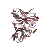
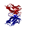
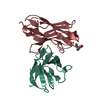
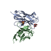
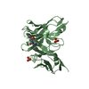
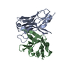
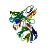
| Title | FV FRAGMENT OF MOUSE MONOCLONAL ANTIBODY D1.3 (BALB/C, IGG1, K) VARIANT CHAIN L GLU81->ASP | ||||||
 Components Components |
| ||||||
 Keywords Keywords | IMMUNOGLOBULIN | ||||||
| Function / homology |  Function and homology information Function and homology informationimmunoglobulin mediated immune response / immunoglobulin complex / antigen binding / adaptive immune response / immune response / extracellular region / plasma membrane Similarity search - Function | ||||||
| Biological species |  | ||||||
| Method |  X-RAY DIFFRACTION / MOLECULAR REPLACEMENT / Resolution: 2.01 Å X-RAY DIFFRACTION / MOLECULAR REPLACEMENT / Resolution: 2.01 Å | ||||||
 Authors Authors | Marks, C. / Henrick, K. / Winter, G. | ||||||
 Citation Citation | Journal: To be Published Title: X-Ray Structures of D1.3 Fv Mutants Authors: Marks, C. / Henrick, K. / Winter, G. #1: Journal: Proc.Natl.Acad.Sci.USA / Year: 1994Title: Bound Water Molecules and Conformational Stabilization Help Mediate an Antigen-Antibody Association Authors: Bhat, T.N. / Bentley, G.A. / Boulot, G. / Greene, M.I. / Tello, D. / Dall'Acqua, W. / Souchon, H. / Schwarz, F.P. / Mariuzza, R.A. / Poljak, R.J. #2: Journal: Nature / Year: 1990Title: Small Rearrangements in Structures of Fv and Fab Fragments of Antibody D1.3 On Antigen Binding Authors: Bhat, T.N. / Bentley, G.A. / Fischmann, T.O. / Boulot, G. / Poljak, R.J. | ||||||
| History |
|
- Structure visualization
| Structure viewer | Molecule: MolmilJmol/JSmol |
|---|
- Downloads & links
-Download
| PDBx/mmCIF format | 1a7r.cif.gz | 58.9 KB | Display | PDBx/mmCIF format |
|---|---|---|---|---|
| PDB format | pdb1a7r.ent.gz | 42.1 KB | Display | PDB format |
| PDBx/mmJSON format | 1a7r.json.gz | Tree view | PDBx/mmJSON format | |
| Others |  Other downloads Other downloads |
-Validation report
| Arichive directory | https://data.pdbj.org/pub/pdb/validation_reports/a7/1a7rftp://data.pdbj.org/pub/pdb/validation_reports/a7/1a7r | HTTPS FTP |
|---|
-Related structure data
| Related structure data | 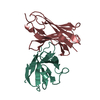 1a7nC  1a7oC 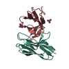 1a7pC  1a7qC 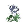 1vfaS S: Starting model for refinement C: citing same article ( |
|---|---|
| Similar structure data |


-Links
 PDBj
PDBj

- Assembly
| Deposited unit | 
| ||||||||
|---|---|---|---|---|---|---|---|---|---|
| 1 |
| ||||||||
| Unit cell |
|
-Components
| #1: Antibody | Mass: 11686.950 Da / Num. of mol.: 1 / Fragment: FV FRAGMENT / Mutation: E81D Source method: isolated from a genetically manipulated source Source: (gene. exp.)  |
|---|---|
| #2: Antibody | Mass: 12857.275 Da / Num. of mol.: 1 / Fragment: FV FRAGMENT Source method: isolated from a genetically manipulated source Source: (gene. exp.) |
| #3: Water | ChemComp-HOH /  Mass: 18.015 Da / Num. of mol.: 111 / Source method: isolated from a natural source / Formula: H2O Mass: 18.015 Da / Num. of mol.: 111 / Source method: isolated from a natural source / Formula: H2O |
| Compound details | THE ANTIBODY IS SECRETED INTO PERIPLASMIC SPACE. VH AND VL DOMAINS ARE COVALENTLY LINKED AND THEY ...THE ANTIBODY IS SECRETED INTO PERIPLASMI |
| Has protein modification | Y |
-Experimental details
-Experiment
| Experiment | Method: X-RAY DIFFRACTION / Number of used crystals: 1 |
|---|
- Sample preparation
| Crystal | Density Matthews: 2.33 Å3/Da / Density % sol: 47.25 % |
|---|
-Data collection
| Diffraction | Mean temperature: 269 K |
|---|---|
| Diffraction source | Wavelength: 1.5418 |
| Detector | Type: MARRESEARCH / Detector: IMAGE PLATE / Date: Dec 1, 1992 |
| Radiation | Monochromatic (M) / Laue (L): M / Scattering type: x-ray |
| Radiation wavelength | Wavelength: 1.5418 Å / Relative weight: 1 |
| Reflection | Resolution: 2→13.89 Å / Num. obs: 96880 / % possible obs: 94.7 % / Observed criterion σ(I): 0 / Redundancy: 4.3 % / Biso Wilson estimate: 15.94 Å2 / Rmerge(I) obs: 0.08 |
| Reflection shell | Resolution: 2→2.05 Å / Redundancy: 2.6 % / Rmerge(I) obs: 0.297 / % possible all: 43.3 |
- Processing
| Software |
| ||||||||||||||||||||||||||||||||||||||||||||||||||||||||||||||||||||||||||||||||||||
|---|---|---|---|---|---|---|---|---|---|---|---|---|---|---|---|---|---|---|---|---|---|---|---|---|---|---|---|---|---|---|---|---|---|---|---|---|---|---|---|---|---|---|---|---|---|---|---|---|---|---|---|---|---|---|---|---|---|---|---|---|---|---|---|---|---|---|---|---|---|---|---|---|---|---|---|---|---|---|---|---|---|---|---|---|---|
| Refinement | Method to determine structure: MOLECULAR REPLACEMENT Starting model: PDB ENTRY 1VFA Resolution: 2.01→6 Å / σ(F): 0 /
| ||||||||||||||||||||||||||||||||||||||||||||||||||||||||||||||||||||||||||||||||||||
| Displacement parameters | Biso mean: 18.25 Å2 | ||||||||||||||||||||||||||||||||||||||||||||||||||||||||||||||||||||||||||||||||||||
| Refinement step | Cycle: LAST / Resolution: 2.01→6 Å
| ||||||||||||||||||||||||||||||||||||||||||||||||||||||||||||||||||||||||||||||||||||
| Refine LS restraints |
|
