 Movie
Movie Controller

[English] 日本語
 Yorodumi
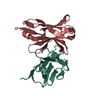
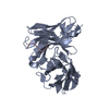
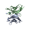
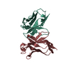
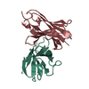
- PDB-1dql: CRYSTAL STRUCTURE OF AN UNLIGANDED (NATIVE) FV FROM A HUMAN IGM A... -
+ Open data

- Basic information
| Entry | Database: PDB / ID: 1dql | |||||||||
|---|---|---|---|---|---|---|---|---|---|---|
| Title | CRYSTAL STRUCTURE OF AN UNLIGANDED (NATIVE) FV FROM A HUMAN IGM ANTI-PEPTIDE ANTIBODY | |||||||||
 Components Components | (IGM MEZ IMMUNOGLOBULIN) x 2 | |||||||||
 Keywords Keywords | IMMUNE SYSTEM / IMMUNOGLOBULIN FOLD / ANTIBODY / IgM / Fv | |||||||||
| Function / homology | Immunoglobulins / Immunoglobulin-like / Sandwich / Mainly Beta Function and homology information Function and homology information | |||||||||
| Biological species |  Homo sapiens (human) Homo sapiens (human) | |||||||||
| Method |  X-RAY DIFFRACTION / Resolution: 2.6 Å X-RAY DIFFRACTION / Resolution: 2.6 Å | |||||||||
 Authors Authors | Ramsland, P.A. / Shan, L. / Moomaw, C.R. / Slaughter, C.A. / Guddat, L.W. / Edmundson, A.B. | |||||||||
 Citation Citation | Journal: Mol.Immunol. / Year: 2000 Title: An unusual human IgM antibody with a protruding HCDR3 and high avidity for its peptide ligands. Authors: Ramsland, P.A. / Shan, L. / Moomaw, C.R. / Slaughter, C.A. / Fan, Z. / Guddat, L.W. / Edmundson, A.B. | |||||||||
| History |
|
- Structure visualization
| Structure viewer | Molecule: MolmilJmol/JSmol |
|---|
- Downloads & links
-Download
| PDBx/mmCIF format | 1dql.cif.gz | 55.9 KB | Display | PDBx/mmCIF format |
|---|---|---|---|---|
| PDB format | pdb1dql.ent.gz | 39.8 KB | Display | PDB format |
| PDBx/mmJSON format | 1dql.json.gz | Tree view | PDBx/mmJSON format | |
| Others |  Other downloads Other downloads |
-Validation report
| Arichive directory | https://data.pdbj.org/pub/pdb/validation_reports/dq/1dqlftp://data.pdbj.org/pub/pdb/validation_reports/dq/1dql | HTTPS FTP |
|---|
-Related structure data
| Similar structure data |
|---|


-Links
 PDBj
PDBj

- Assembly
| Deposited unit | 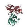
| ||||||||
|---|---|---|---|---|---|---|---|---|---|
| 1 |
| ||||||||
| Unit cell |
| ||||||||
| Details | The Mez Fv is a non-covalent heterodimer of VL (chain L) and VH (chain H) |
-Components
| #1: Antibody | Mass: 11527.743 Da / Num. of mol.: 1 / Fragment: LIGHT CHAIN VARIABLE DOMAIN FRAGMENT / Source method: isolated from a natural source / Source: (natural) Homo sapiens (human) |
|---|---|
| #2: Antibody | Mass: 13214.604 Da / Num. of mol.: 1 / Fragment: HEAVY CHAIN VARIABLE DOMAIN FRAGMENT / Source method: isolated from a natural source / Source: (natural) Homo sapiens (human) |
| #3: Water | ChemComp-HOH /  Mass: 18.015 Da / Num. of mol.: 18 / Source method: isolated from a natural source / Formula: H2O Mass: 18.015 Da / Num. of mol.: 18 / Source method: isolated from a natural source / Formula: H2O |
| Has protein modification | Y |
-Experimental details
-Experiment
| Experiment | Method: X-RAY DIFFRACTION / Number of used crystals: 1 |
|---|
- Sample preparation
| Crystal | Density Matthews: 2.72 Å3/Da / Density % sol: 55.2 % | ||||||||||||||||||||
|---|---|---|---|---|---|---|---|---|---|---|---|---|---|---|---|---|---|---|---|---|---|
| Crystal grow | Temperature: 295 K / Method: microseeding, sitting drop / pH: 7 Details: 6-8% (W/V) PEG 8000, 0.05% (W/V) SODIUM AZIDE, pH 7.0, MICROSEEDING, SITTING DROP, temperature 295K | ||||||||||||||||||||
| Crystal grow | *PLUS Method: unknown | ||||||||||||||||||||
| Components of the solutions | *PLUS
|
-Data collection
| Diffraction | Mean temperature: 288 K |
|---|---|
| Diffraction source | Source: ROTATING ANODE / Type: SIEMENS / Wavelength: 1.5418 |
| Detector | Type: SIEMENS / Detector: AREA DETECTOR / Date: Nov 19, 1990 |
| Radiation | Protocol: SINGLE WAVELENGTH / Monochromatic (M) / Laue (L): M / Scattering type: x-ray |
| Radiation wavelength | Wavelength: 1.5418 Å / Relative weight: 1 |
| Reflection | Resolution: 2.58→20 Å / Num. all: 7111 / Num. obs: 7111 / % possible obs: 84.4 % / Observed criterion σ(F): 0 / Observed criterion σ(I): 0 / Redundancy: 2.25 % / Biso Wilson estimate: 13.1 Å2 / Rmerge(I) obs: 0.145 / Net I/σ(I): 12.4 |
| Reflection shell | Resolution: 2.58→2.75 Å / Redundancy: 1.2 % / Rmerge(I) obs: 0.167 / % possible all: 40 |
| Reflection | *PLUS Highest resolution: 2.6 Å / % possible obs: 83.4 % |
| Reflection shell | *PLUS Highest resolution: 2.6 Å / Lowest resolution: 2.76 Å / % possible obs: 40.6 % |
- Processing
| Software |
| ||||||||||||||||||||||||||||||||||||||||||||||||||||||||||||
|---|---|---|---|---|---|---|---|---|---|---|---|---|---|---|---|---|---|---|---|---|---|---|---|---|---|---|---|---|---|---|---|---|---|---|---|---|---|---|---|---|---|---|---|---|---|---|---|---|---|---|---|---|---|---|---|---|---|---|---|---|---|
| Refinement | Resolution: 2.6→20 Å / Rfactor Rfree error: 0.009 / Cross valid method: THROUGHOUT / σ(F): 0 / σ(I): 0 / Stereochemistry target values: ENGH & HUBER
| ||||||||||||||||||||||||||||||||||||||||||||||||||||||||||||
| Solvent computation | Solvent model: FLAT MODEL / Bsol: 14.31 Å2 / ksol: 0.252 e/Å3 | ||||||||||||||||||||||||||||||||||||||||||||||||||||||||||||
| Displacement parameters | Biso mean: 18.9 Å2 | ||||||||||||||||||||||||||||||||||||||||||||||||||||||||||||
| Refine analyze |
| ||||||||||||||||||||||||||||||||||||||||||||||||||||||||||||
| Refinement step | Cycle: LAST / Resolution: 2.6→20 Å
| ||||||||||||||||||||||||||||||||||||||||||||||||||||||||||||
| Refine LS restraints |
| ||||||||||||||||||||||||||||||||||||||||||||||||||||||||||||
| LS refinement shell | Resolution: 2.6→2.76 Å / Rfactor Rfree error: 0.047 / Total num. of bins used: 6
| ||||||||||||||||||||||||||||||||||||||||||||||||||||||||||||
| Xplor file |
| ||||||||||||||||||||||||||||||||||||||||||||||||||||||||||||
| Software | *PLUS Name: CNS / Classification: refinement | ||||||||||||||||||||||||||||||||||||||||||||||||||||||||||||
| Refine LS restraints | *PLUS
|
