 Movie
Movie Controller


+ Open data

- Basic information
| Entry | Database: PDB / ID: 1a4e | ||||||
|---|---|---|---|---|---|---|---|
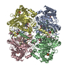
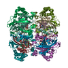
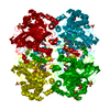
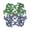
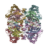
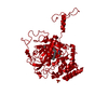
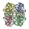
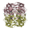
| Title | CATALASE A FROM SACCHAROMYCES CEREVISIAE | ||||||
 Components Components | CATALASE A | ||||||
 Keywords Keywords | OXIDOREDUCTASE / CATALASE / PEROXIDASE | ||||||
| Function / homology |  Function and homology information Function and homology informationPeroxisomal protein import / Detoxification of Reactive Oxygen Species / catalase / catalase activity / peroxisomal matrix / Neutrophil degranulation / hydrogen peroxide catabolic process / response to reactive oxygen species / response to hydrogen peroxide / peroxisome ...Peroxisomal protein import / Detoxification of Reactive Oxygen Species / catalase / catalase activity / peroxisomal matrix / Neutrophil degranulation / hydrogen peroxide catabolic process / response to reactive oxygen species / response to hydrogen peroxide / peroxisome / mitochondrial matrix / heme binding / mitochondrion / metal ion binding / cytoplasm Similarity search - Function | ||||||
| Biological species |  | ||||||
| Method |  X-RAY DIFFRACTION / SYNCHROTRON / MOLECULAR REPLACEMENT / Resolution: 2.4 Å X-RAY DIFFRACTION / SYNCHROTRON / MOLECULAR REPLACEMENT / Resolution: 2.4 Å | ||||||
 Authors Authors | Mate, M.J. | ||||||
 Citation Citation | Journal: J.Mol.Biol. / Year: 1999 Title: Structure of catalase-A from Saccharomyces cerevisiae. Authors: Mate, M.J. / Zamocky, M. / Nykyri, L.M. / Herzog, C. / Alzari, P.M. / Betzel, C. / Koller, F. / Fita, I. | ||||||
| History |
|
- Structure visualization
| Structure viewer | Molecule: MolmilJmol/JSmol |
|---|
- Downloads & links
-Download
| PDBx/mmCIF format | 1a4e.cif.gz | 415.4 KB | Display | PDBx/mmCIF format |
|---|---|---|---|---|
| PDB format | pdb1a4e.ent.gz | 337.7 KB | Display | PDB format |
| PDBx/mmJSON format | 1a4e.json.gz | Tree view | PDBx/mmJSON format | |
| Others |  Other downloads Other downloads |
-Validation report
| Arichive directory | https://data.pdbj.org/pub/pdb/validation_reports/a4/1a4eftp://data.pdbj.org/pub/pdb/validation_reports/a4/1a4e | HTTPS FTP |
|---|
-Related structure data
| Related structure data |  7catS S: Starting model for refinement |
|---|---|
| Similar structure data |


-Links
 PDBj
PDBj


- Assembly
| Deposited unit | 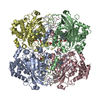
| ||||||||||||||||
|---|---|---|---|---|---|---|---|---|---|---|---|---|---|---|---|---|---|
| 1 |
| ||||||||||||||||
| Unit cell |
| ||||||||||||||||
| Noncrystallographic symmetry (NCS) | NCS oper:
|
-Components
| #1: Protein | Mass: 55623.930 Da / Num. of mol.: 4 / Source method: isolated from a natural source / Source: (natural) #2: Chemical | ChemComp-AZI /   Mass: 42.020 Da / Num. of mol.: 4 / Source method: obtained synthetically / Formula: N3 Mass: 42.020 Da / Num. of mol.: 4 / Source method: obtained synthetically / Formula: N3#3: Chemical |   Mass: 96.063 Da / Num. of mol.: 2 / Source method: obtained synthetically / Formula: SO4 Mass: 96.063 Da / Num. of mol.: 2 / Source method: obtained synthetically / Formula: SO4#4: Chemical | ChemComp-HEM /   Mass: 616.487 Da / Num. of mol.: 4 / Source method: obtained synthetically / Formula: C34H32FeN4O4 Mass: 616.487 Da / Num. of mol.: 4 / Source method: obtained synthetically / Formula: C34H32FeN4O4#5: Water | ChemComp-HOH / |  Mass: 18.015 Da / Num. of mol.: 957 / Source method: isolated from a natural source / Formula: H2O Mass: 18.015 Da / Num. of mol.: 957 / Source method: isolated from a natural source / Formula: H2O |
|---|
-Experimental details
-Experiment
| Experiment | Method: X-RAY DIFFRACTION / Number of used crystals: 1 |
|---|
- Sample preparation
| Crystal | Density Matthews: 3.35 Å3/Da / Density % sol: 60 % | ||||||||||||||||||||
|---|---|---|---|---|---|---|---|---|---|---|---|---|---|---|---|---|---|---|---|---|---|
| Crystal grow | pH: 8.5 / Details: pH 8.5 | ||||||||||||||||||||
| Crystal | *PLUS | ||||||||||||||||||||
| Crystal grow | *PLUS Method: vapor diffusion, hanging drop | ||||||||||||||||||||
| Components of the solutions | *PLUS
|
-Data collection
| Diffraction | Mean temperature: 293 K |
|---|---|
| Diffraction source | Source: SYNCHROTRON / Site: EMBL/DESY, HAMBURG  / Type: EMBL/DESY, HAMBURG / Wavelength: 0.928 / Type: EMBL/DESY, HAMBURG / Wavelength: 0.928 |
| Detector | Type: MARRESEARCH / Detector: IMAGE PLATE / Date: Feb 1, 1995 |
| Radiation | Monochromatic (M) / Laue (L): M / Scattering type: x-ray |
| Radiation wavelength | Wavelength: 0.928 Å / Relative weight: 1 |
| Reflection | Resolution: 2.35→30 Å / Num. obs: 123230 / % possible obs: 97.7 % / Observed criterion σ(I): 2 / Biso Wilson estimate: 33.6 Å2 / Rmerge(I) obs: 0.074 / Rsym value: 0.074 / Net I/σ(I): 25.12 |
| Reflection shell | Resolution: 2.35→2.39 Å / Rmerge(I) obs: 0.33 / Mean I/σ(I) obs: 5.69 / Rsym value: 0.33 / % possible all: 94.5 |
| Reflection | *PLUS Num. measured all: 1276062 |
| Reflection shell | *PLUS % possible obs: 95 % |
- Processing
| Software |
| ||||||||||||||||||||||||||||||||||||||||||||||||||||||||||||||||||||||||||||||||
|---|---|---|---|---|---|---|---|---|---|---|---|---|---|---|---|---|---|---|---|---|---|---|---|---|---|---|---|---|---|---|---|---|---|---|---|---|---|---|---|---|---|---|---|---|---|---|---|---|---|---|---|---|---|---|---|---|---|---|---|---|---|---|---|---|---|---|---|---|---|---|---|---|---|---|---|---|---|---|---|---|---|
| Refinement | Method to determine structure: MOLECULAR REPLACEMENT Starting model: PDB ENTRY 7CAT Resolution: 2.4→20 Å / Rfactor Rfree error: 0.002 / Data cutoff high absF: 10000000 / Data cutoff low absF: 0.001 / Cross valid method: THROUGHOUT / σ(F): 0
| ||||||||||||||||||||||||||||||||||||||||||||||||||||||||||||||||||||||||||||||||
| Displacement parameters | Biso mean: 33.9 Å2 | ||||||||||||||||||||||||||||||||||||||||||||||||||||||||||||||||||||||||||||||||
| Refine analyze |
| ||||||||||||||||||||||||||||||||||||||||||||||||||||||||||||||||||||||||||||||||
| Refinement step | Cycle: LAST / Resolution: 2.4→20 Å
| ||||||||||||||||||||||||||||||||||||||||||||||||||||||||||||||||||||||||||||||||
| Refine LS restraints |
| ||||||||||||||||||||||||||||||||||||||||||||||||||||||||||||||||||||||||||||||||
| LS refinement shell | Resolution: 2.4→2.49 Å / Rfactor Rfree error: 0.008 / Total num. of bins used: 10
| ||||||||||||||||||||||||||||||||||||||||||||||||||||||||||||||||||||||||||||||||
| Software | *PLUS Name: X-PLOR / Version: 3.851 / Classification: refinement | ||||||||||||||||||||||||||||||||||||||||||||||||||||||||||||||||||||||||||||||||
| Refine LS restraints | *PLUS
|
