 Movie
Movie Controller

[English] 日本語
 Yorodumi
- PDB-1h7k: Formation of a tyrosyl radical intermediate in Proteus mirabilis ... -
+ Open data

- Basic information
| Entry | Database: PDB / ID: 1h7k | ||||||
|---|---|---|---|---|---|---|---|
| Title | Formation of a tyrosyl radical intermediate in Proteus mirabilis catalase by directed mutagenesis and consequences for nucleotide reactivity | ||||||
 Components Components | CATALASE | ||||||
 Keywords Keywords | OXIDOREDUCTASE / OXIDOREDUCTASE (H2O2 ACCEPTOR) / PEROXIDASE / IRON / HEM / HYDROGEN PEROXIDE / NADP | ||||||
| Function / homology |  Function and homology information Function and homology informationcatalase / catalase activity / hydrogen peroxide catabolic process / response to hydrogen peroxide / heme binding / metal ion binding / cytoplasm Similarity search - Function | ||||||
| Biological species |  PROTEUS MIRABILIS (bacteria) PROTEUS MIRABILIS (bacteria) | ||||||
| Method |  X-RAY DIFFRACTION / SYNCHROTRON / MOLECULAR REPLACEMENT / Resolution: 2.4 Å X-RAY DIFFRACTION / SYNCHROTRON / MOLECULAR REPLACEMENT / Resolution: 2.4 Å | ||||||
 Authors Authors | Andreoletti, P. / Gambarelli, S. / Gaillard, J. / Sainz, G. / Stojanoff, V. / Jouve, H.M. | ||||||
 Citation Citation | Journal: Biochemistry / Year: 2001 Title: Formation of a Tyrosyl Radical Intermediate in Proteus Mirabilis Catalase by Directed Mutagenesis and Consequences for Nucleotide Reactivity. Authors: Andreoletti, P. / Gambarelli, S. / Sainz, G. / Stojanoff, V. / White, C. / Desfonds, G. / Gagnon, J. / Gaillard, J. / Jouve, H.M. #1: Journal: Nat.Struct.Biol. / Year: 1996Title: Ferryl Intermediates of Catalase Captured by Time-Resolved Weissenberg Crystallography and Uv-Vis Spectroscopy Authors: Gouet, P. / Jouve, H.M. / Williams, P.A. / Andersson, I. / Andreoletti, P. / Nussaume, L. / Hajdu, J. #2: Journal: J.Mol.Biol. / Year: 1995Title: Crystal Structure of Proteus Mirabilis Pr Catalase with and without Bound Nadph Authors: Gouet, P. / Jouve, H.M. / Dideberg, O. #3: Journal: J.Mol.Biol. / Year: 1991 Title: Crystallization and Crystal Packing of Proteus Mirabilis Pr Catalase Authors: Jouve, H.M. / Gouet, P. / Boudjada, N. / Buisson, G. / Kahn, R. / Duee, E. #4: Journal: Acta Crystallogr.,Sect.B / Year: 1986Title: The Refined Structure of Beef Liver Catalase at 2.5 Angstroms Resolution Authors: Fita, I. / Silva, A.M. / Murthy, M.R.N. / Rossmann, M.G. | ||||||
| History |
| ||||||
| Remark 700 | SHEET DETERMINATION METHOD: DSSP THE SHEETS PRESENTED AS "AA" IN EACH CHAIN ON SHEET RECORDS BELOW ... SHEET DETERMINATION METHOD: DSSP THE SHEETS PRESENTED AS "AA" IN EACH CHAIN ON SHEET RECORDS BELOW IS ACTUALLY AN 10-STRANDED BARREL THIS IS REPRESENTED BY A 11-STRANDED SHEET IN WHICH THE FIRST AND LAST STRANDS ARE IDENTICAL. |
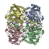
- Structure visualization
| Structure viewer | Molecule: MolmilJmol/JSmol |
|---|
- Downloads & links
-Download
| PDBx/mmCIF format | 1h7k.cif.gz | 116.4 KB | Display | PDBx/mmCIF format |
|---|---|---|---|---|
| PDB format | pdb1h7k.ent.gz | 88 KB | Display | PDB format |
| PDBx/mmJSON format | 1h7k.json.gz | Tree view | PDBx/mmJSON format | |
| Others |  Other downloads Other downloads |
-Validation report
| Arichive directory | https://data.pdbj.org/pub/pdb/validation_reports/h7/1h7kftp://data.pdbj.org/pub/pdb/validation_reports/h7/1h7k | HTTPS FTP |
|---|
-Related structure data
| Related structure data |  1cae S: Starting model for refinement |
|---|---|
| Similar structure data |

-Links
 PDBj
PDBj
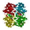
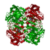
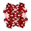
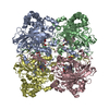
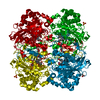
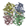
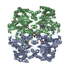
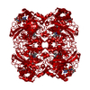
- Assembly
| Deposited unit | 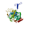
| ||||||||
|---|---|---|---|---|---|---|---|---|---|
| 1 | 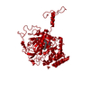
| ||||||||
| Unit cell |
| ||||||||
| Components on special symmetry positions |
|
-Components
| #1: Protein | Mass: 55611.152 Da / Num. of mol.: 1 Source method: isolated from a genetically manipulated source Details: METHIONINE SULFONE IN POSITION 53, TYROSINE 337 LACK THE HYDROXYL HYDROGEN Source: (gene. exp.) PROTEUS MIRABILIS (bacteria) / Plasmid: PALTER-CAT-F215Y / Production host: |
|---|---|
| #2: Chemical | ChemComp-HEM /   Mass: 616.487 Da / Num. of mol.: 1 / Source method: obtained synthetically / Formula: C34H32FeN4O4 Mass: 616.487 Da / Num. of mol.: 1 / Source method: obtained synthetically / Formula: C34H32FeN4O4 |
| #3: Chemical | ChemComp-ACT /   Mass: 59.044 Da / Num. of mol.: 1 / Source method: obtained synthetically / Formula: C2H3O2 Mass: 59.044 Da / Num. of mol.: 1 / Source method: obtained synthetically / Formula: C2H3O2 |
| #4: Chemical | ChemComp-SO4 /   Mass: 96.063 Da / Num. of mol.: 1 / Source method: obtained synthetically / Formula: SO4 Mass: 96.063 Da / Num. of mol.: 1 / Source method: obtained synthetically / Formula: SO4 |
| #5: Water | ChemComp-HOH /  Mass: 18.015 Da / Num. of mol.: 168 / Source method: isolated from a natural source / Formula: H2O Mass: 18.015 Da / Num. of mol.: 168 / Source method: isolated from a natural source / Formula: H2O |
| Has protein modification | Y |
| Sequence details | MODRES: 1H7K OMT A 53() METHIONINE SULFONE (S-DIOXYMETHIONINE) MODRES: 1H7K TYR A 337() OXYGEN OF ...MODRES: 1H7K OMT A 53() METHIONINE |
-Experimental details
-Experiment
| Experiment | Method: X-RAY DIFFRACTION / Number of used crystals: 1 |
|---|
- Sample preparation
| Crystal | Density Matthews: 3.82 Å3/Da / Density % sol: 63 % | ||||||||||||||||||||||||||||||||||||||||||||||||||||||
|---|---|---|---|---|---|---|---|---|---|---|---|---|---|---|---|---|---|---|---|---|---|---|---|---|---|---|---|---|---|---|---|---|---|---|---|---|---|---|---|---|---|---|---|---|---|---|---|---|---|---|---|---|---|---|---|
| Crystal grow | Temperature: 277 K / Method: vapor diffusion, hanging drop / pH: 7.3 / Details: HANGING DROP AT 4 DEG C, pH 7.30 | ||||||||||||||||||||||||||||||||||||||||||||||||||||||
| Crystal grow | *PLUS Temperature: 4-5 ℃ / pH: 7.5 / Method: vapor diffusion, hanging drop / Details: Jouve, H.M., (1991) J.Mol.Biol., 221, 1075. | ||||||||||||||||||||||||||||||||||||||||||||||||||||||
| Components of the solutions | *PLUS
|
-Data collection
| Diffraction | Mean temperature: 100 K |
|---|---|
| Diffraction source | Source: SYNCHROTRON / Site: ESRF  / Beamline: ID14-4 / Wavelength: 0.9574 / Beamline: ID14-4 / Wavelength: 0.9574 |
| Detector | Type: MARRESEARCH / Detector: IMAGE PLATE / Date: Jul 15, 1998 |
| Radiation | Protocol: SINGLE WAVELENGTH / Monochromatic (M) / Laue (L): M / Scattering type: x-ray |
| Radiation wavelength | Wavelength: 0.9574 Å / Relative weight: 1 |
| Reflection | Resolution: 2.4→29.26 Å / Num. obs: 35932 / % possible obs: 99.7 % / Observed criterion σ(I): 3 / Redundancy: 15.8 % / Biso Wilson estimate: 40 Å2 |
| Reflection shell | Resolution: 2→2.05 Å / % possible all: 94.6 |
| Reflection | *PLUS Redundancy: 15.8 % / Rmerge(I) obs: 0.08 |
| Reflection shell | *PLUS % possible obs: 99.9 % / Redundancy: 4.8 % / Rmerge(I) obs: 0.251 |
- Processing
| Software |
| ||||||||||||||||||||||||||||||||||||||||||||||||||||||||||||||||||||||||||||||||
|---|---|---|---|---|---|---|---|---|---|---|---|---|---|---|---|---|---|---|---|---|---|---|---|---|---|---|---|---|---|---|---|---|---|---|---|---|---|---|---|---|---|---|---|---|---|---|---|---|---|---|---|---|---|---|---|---|---|---|---|---|---|---|---|---|---|---|---|---|---|---|---|---|---|---|---|---|---|---|---|---|---|
| Refinement | Method to determine structure: MOLECULAR REPLACEMENT Starting model: PDB ENTRY 1CAE 1cae Resolution: 2.4→29.26 Å / Rfactor Rfree error: 0.005 / Data cutoff high absF: 2727867.74 / Isotropic thermal model: RESTRAINED / Cross valid method: THROUGHOUT / σ(F): 0 / Details: AMINO ACIDS 358 - 362 ARE NOT REFINED
| ||||||||||||||||||||||||||||||||||||||||||||||||||||||||||||||||||||||||||||||||
| Solvent computation | Solvent model: FLAT MODEL / Bsol: 40.7587 Å2 / ksol: 0.306189 e/Å3 | ||||||||||||||||||||||||||||||||||||||||||||||||||||||||||||||||||||||||||||||||
| Displacement parameters | Biso mean: 51.2 Å2
| ||||||||||||||||||||||||||||||||||||||||||||||||||||||||||||||||||||||||||||||||
| Refine analyze |
| ||||||||||||||||||||||||||||||||||||||||||||||||||||||||||||||||||||||||||||||||
| Refinement step | Cycle: LAST / Resolution: 2.4→29.26 Å
| ||||||||||||||||||||||||||||||||||||||||||||||||||||||||||||||||||||||||||||||||
| Refine LS restraints |
| ||||||||||||||||||||||||||||||||||||||||||||||||||||||||||||||||||||||||||||||||
| LS refinement shell | Resolution: 2→2.12 Å / Rfactor Rfree error: 0.013 / Total num. of bins used: 6
| ||||||||||||||||||||||||||||||||||||||||||||||||||||||||||||||||||||||||||||||||
| Xplor file |
| ||||||||||||||||||||||||||||||||||||||||||||||||||||||||||||||||||||||||||||||||
| Refinement | *PLUS Lowest resolution: 15 Å / % reflection Rfree: 5 % / Rfactor Rfree: 0.24 | ||||||||||||||||||||||||||||||||||||||||||||||||||||||||||||||||||||||||||||||||
| Solvent computation | *PLUS | ||||||||||||||||||||||||||||||||||||||||||||||||||||||||||||||||||||||||||||||||
| Displacement parameters | *PLUS | ||||||||||||||||||||||||||||||||||||||||||||||||||||||||||||||||||||||||||||||||
| Refine LS restraints | *PLUS
|
