 Movie
Movie Controller

[English] 日本語
 Yorodumi
- PDB-1ye9: Crystal structure of proteolytically truncated catalase HPII from... -
+ Open data

- Basic information
| Entry | Database: PDB / ID: 1ye9 | ||||||
|---|---|---|---|---|---|---|---|
| Title | Crystal structure of proteolytically truncated catalase HPII from E. coli | ||||||
 Components Components | (catalase HPII) x 2 | ||||||
 Keywords Keywords | OXIDOREDUCTASE / catalase HPII / proteolytic truncation / beta barrel core | ||||||
| Function / homology |  Function and homology information Function and homology informationcatalase / hyperosmotic response / catalase activity / hydrogen peroxide catabolic process / response to oxidative stress / iron ion binding / heme binding / DNA damage response / identical protein binding / cytosol / cytoplasm Similarity search - Function | ||||||
| Biological species |  | ||||||
| Method |  X-RAY DIFFRACTION / SYNCHROTRON / MOLECULAR REPLACEMENT / Resolution: 2.8 Å X-RAY DIFFRACTION / SYNCHROTRON / MOLECULAR REPLACEMENT / Resolution: 2.8 Å | ||||||
 Authors Authors | Loewen, P.C. / Chelikani, P. / Carpena, X. / Fita, I. / Perez-Luque, R. / Donald, L.J. / Switala, J. / Duckworth, H.W. | ||||||
 Citation Citation | Journal: Biochemistry / Year: 2005 Title: Characterization of a Large Subunit Catalase Truncated by Proteolytic Cleavage(,) Authors: Chelikani, P. / Carpena, X. / Perez-Luque, R. / Donald, L.J. / Duckworth, H.W. / Switala, J. / Fita, I. / Loewen, P.C. | ||||||
| History |
|
- Structure visualization
| Structure viewer | Molecule: MolmilJmol/JSmol |
|---|
- Downloads & links
-Download
| PDBx/mmCIF format | 1ye9.cif.gz | 755.4 KB | Display | PDBx/mmCIF format |
|---|---|---|---|---|
| PDB format | pdb1ye9.ent.gz | 627.6 KB | Display | PDB format |
| PDBx/mmJSON format | 1ye9.json.gz | Tree view | PDBx/mmJSON format | |
| Others |  Other downloads Other downloads |
-Validation report
| Arichive directory | https://data.pdbj.org/pub/pdb/validation_reports/ye/1ye9ftp://data.pdbj.org/pub/pdb/validation_reports/ye/1ye9 | HTTPS FTP |
|---|
-Related structure data
| Related structure data |  1ggeS S: Starting model for refinement |
|---|---|
| Similar structure data |


-Links
 PDBj
PDBj

- Assembly
| Deposited unit | 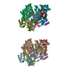
| ||||||||||||||||||||||||||||||||||||||||||||||||||||||
|---|---|---|---|---|---|---|---|---|---|---|---|---|---|---|---|---|---|---|---|---|---|---|---|---|---|---|---|---|---|---|---|---|---|---|---|---|---|---|---|---|---|---|---|---|---|---|---|---|---|---|---|---|---|---|---|
| 1 | 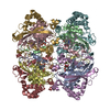
| ||||||||||||||||||||||||||||||||||||||||||||||||||||||
| 2 | 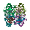
| ||||||||||||||||||||||||||||||||||||||||||||||||||||||
| Unit cell |
| ||||||||||||||||||||||||||||||||||||||||||||||||||||||
| Noncrystallographic symmetry (NCS) | NCS domain:
NCS domain segments: Component-ID: 1 / Ens-ID: 1 / Beg auth comp-ID: SER / Beg label comp-ID: SER / End auth comp-ID: ILE / End label comp-ID: ILE / Refine code: 1 / Auth seq-ID: 75 - 564 / Label seq-ID: 1 - 256
| ||||||||||||||||||||||||||||||||||||||||||||||||||||||
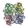
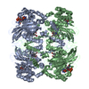
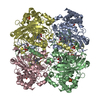
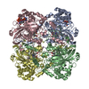
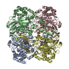
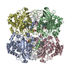
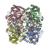
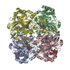
| Details | Biological unit is a tetramer and the asymmetric unit is composed of two truncated tetramers in which each monomer is cut into two fragments. |
-Components
| #1: Protein | Mass: 25615.742 Da / Num. of mol.: 8 / Fragment: proteolytic fragment, residues 75-300 Source method: isolated from a genetically manipulated source Source: (gene. exp.) #2: Protein | Mass: 30072.516 Da / Num. of mol.: 8 / Fragment: proteolytic fragment, residues 309-567 Source method: isolated from a genetically manipulated source Source: (gene. exp.) #3: Chemical | ChemComp-HDD /   Mass: 632.487 Da / Num. of mol.: 8 / Source method: obtained synthetically / Formula: C34H32FeN4O5 Mass: 632.487 Da / Num. of mol.: 8 / Source method: obtained synthetically / Formula: C34H32FeN4O5#4: Water | ChemComp-HOH / |  Mass: 18.015 Da / Num. of mol.: 180 / Source method: isolated from a natural source / Formula: H2O Mass: 18.015 Da / Num. of mol.: 180 / Source method: isolated from a natural source / Formula: H2O |
|---|
-Experimental details
-Experiment
| Experiment | Method: X-RAY DIFFRACTION / Number of used crystals: 1 |
|---|
- Sample preparation
| Crystal | Density Matthews: 2.56 Å3/Da / Density % sol: 51.6 % |
|---|---|
| Crystal grow | Temperature: 298 K / Method: vapor diffusion, hanging drop / pH: 7 Details: 50mM TrisHCl, 8% PEG 20000, 8% PEG MME 550, 0.2M KSCN, 0.1M dithiothreitol, pH 7.0, VAPOR DIFFUSION, HANGING DROP, temperature 298K |
-Data collection
| Diffraction | Mean temperature: 100 K |
|---|---|
| Diffraction source | Source: SYNCHROTRON / Site: ESRF  / Beamline: ID14-3 / Wavelength: 0.931 Å / Beamline: ID14-3 / Wavelength: 0.931 Å |
| Detector | Type: MARRESEARCH / Detector: CCD / Date: Sep 18, 2003 / Details: Mirrors |
| Radiation | Monochromator: Diamond(111), GE(220) / Protocol: SINGLE WAVELENGTH / Monochromatic (M) / Laue (L): M / Scattering type: x-ray |
| Radiation wavelength | Wavelength: 0.931 Å / Relative weight: 1 |
| Reflection | Resolution: 2.8→30 Å / Num. all: 109986 / Num. obs: 104927 / % possible obs: 95.4 % / Observed criterion σ(F): 3 / Observed criterion σ(I): 3 / Redundancy: 3.3 % / Rmerge(I) obs: 0.07 / Rsym value: 0.106 / Net I/σ(I): 6.6 |
| Reflection shell | Resolution: 2.8→2.87 Å / Redundancy: 3 % / Mean I/σ(I) obs: 1 / Num. unique all: 7510 / Rsym value: 0.65 / % possible all: 92.1 |
- Processing
| Software |
| |||||||||||||||||||||||||||||||||||||||||||||||||||||||||||||||||||||||||||||||||||||
|---|---|---|---|---|---|---|---|---|---|---|---|---|---|---|---|---|---|---|---|---|---|---|---|---|---|---|---|---|---|---|---|---|---|---|---|---|---|---|---|---|---|---|---|---|---|---|---|---|---|---|---|---|---|---|---|---|---|---|---|---|---|---|---|---|---|---|---|---|---|---|---|---|---|---|---|---|---|---|---|---|---|---|---|---|---|---|
| Refinement | Method to determine structure: MOLECULAR REPLACEMENT Starting model: PDB Entry 1GGE Resolution: 2.8→30 Å / Cor.coef. Fo:Fc: 0.914 / Cor.coef. Fo:Fc free: 0.865 / SU B: 20.105 / SU ML: 0.378 / Cross valid method: THROUGHOUT / σ(F): 3 / σ(I): 3 / ESU R Free: 0.432 / Stereochemistry target values: MAXIMUM LIKELIHOOD
| |||||||||||||||||||||||||||||||||||||||||||||||||||||||||||||||||||||||||||||||||||||
| Solvent computation | Ion probe radii: 0.8 Å / Shrinkage radii: 0.8 Å / VDW probe radii: 1.4 Å / Solvent model: BABINET MODEL WITH MASK | |||||||||||||||||||||||||||||||||||||||||||||||||||||||||||||||||||||||||||||||||||||
| Displacement parameters | Biso mean: 43.725 Å2
| |||||||||||||||||||||||||||||||||||||||||||||||||||||||||||||||||||||||||||||||||||||
| Refinement step | Cycle: LAST / Resolution: 2.8→30 Å
| |||||||||||||||||||||||||||||||||||||||||||||||||||||||||||||||||||||||||||||||||||||
| Refine LS restraints |
| |||||||||||||||||||||||||||||||||||||||||||||||||||||||||||||||||||||||||||||||||||||
| Refine LS restraints NCS | Ens-ID: 1 / Number: 3851 / Refine-ID: X-RAY DIFFRACTION
| |||||||||||||||||||||||||||||||||||||||||||||||||||||||||||||||||||||||||||||||||||||
| LS refinement shell | Resolution: 2.8→2.872 Å / Total num. of bins used: 20 /
|
