 Movie
Movie Controller

[English] 日本語
 Yorodumi
- PDB-134d: SOLUTION STRUCTURE OF A PURINE(DOT)PURINE(DOT)PYRIMIDINE DNA TRIP... -
+ Open data

- Basic information
| Entry | Database: PDB / ID: 134d | ||||||
|---|---|---|---|---|---|---|---|
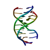
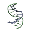
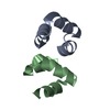
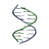
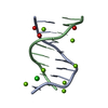
| Title | SOLUTION STRUCTURE OF A PURINE(DOT)PURINE(DOT)PYRIMIDINE DNA TRIPLEX CONTAINING G(DOT)GC AND T(DOT)AT TRIPLES | ||||||
 Components Components | DNA TRIPLEX | ||||||
 Keywords Keywords | DNA / TRIPLEX | ||||||
| Function / homology | DNA / DNA (> 10) Function and homology information Function and homology information | ||||||
| Biological species | synthetic construct (others) | ||||||
| Method | SOLUTION NMR / RESTRAINED MOLECULAR DYNAMICS, DISTANCE GEOMETRY | ||||||
 Authors Authors | Patel, D.J. / Radhakrishnan, I. | ||||||
 Citation Citation | Journal: Structure / Year: 1993 Title: Solution structure of a purine.purine.pyrimidine DNA triplex containing G.GC and T.AT triples. Authors: Radhakrishnan, I. / Patel, D.J. #1: Journal: J.Am.Chem.Soc. / Year: 1993Title: Solution/structure of an intramolecular purine-purine-pyrimidine DNA triplex. Authors: Radhakrishnan, I. / Patel, D.J. | ||||||
| History |
|
- Structure visualization
| Structure viewer | Molecule: MolmilJmol/JSmol |
|---|
- Downloads & links
-Download
| PDBx/mmCIF format | 134d.cif.gz | 26.4 KB | Display | PDBx/mmCIF format |
|---|---|---|---|---|
| PDB format | pdb134d.ent.gz | 17.5 KB | Display | PDB format |
| PDBx/mmJSON format | 134d.json.gz | Tree view | PDBx/mmJSON format | |
| Others |  Other downloads Other downloads |
-Validation report
| Arichive directory | https://data.pdbj.org/pub/pdb/validation_reports/34/134dftp://data.pdbj.org/pub/pdb/validation_reports/34/134d | HTTPS FTP |
|---|
-Related structure data


-Links
 PDBj
PDBj


- Assembly
| Deposited unit | 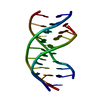
| |||||||||
|---|---|---|---|---|---|---|---|---|---|---|
| 1 |
| |||||||||
| NMR ensembles |
|
-Components
| #1: DNA chain | Mass: 9552.122 Da / Num. of mol.: 1 / Source method: obtained synthetically / Source: (synth.) synthetic construct (others) / Keywords: DEOXYRIBONUCLEIC ACID |
|---|
-Experimental details
-Experiment
| Experiment | Method: SOLUTION NMR |
|---|
- Processing
| Software |
| |||||||||
|---|---|---|---|---|---|---|---|---|---|---|
| NMR software | Name:  X-PLOR / Developer: BRUNGER / Classification: refinement X-PLOR / Developer: BRUNGER / Classification: refinement | |||||||||
| Refinement | Method: RESTRAINED MOLECULAR DYNAMICS, DISTANCE GEOMETRY / Software ordinal: 1 Details: RESTRAINED MOLECULAR DYNAMICS CALCULATIONS WERE DONE ONIDEALIZED A'- AND B-FORM STARTING STRUCTURES. ONLY THE TRIPLEX REGION AND THE FIRST AND LAST RESIDUES OF EACH OF THE TWO LOOPS IN THE ...Details: RESTRAINED MOLECULAR DYNAMICS CALCULATIONS WERE DONE ONIDEALIZED A'- AND B-FORM STARTING STRUCTURES. ONLY THE TRIPLEX REGION AND THE FIRST AND LAST RESIDUES OF EACH OF THE TWO LOOPS IN THE SEQUENCE WERE CONSIDERED. THE REFINEMENT WAS CONDUCTED IN TWO STAGES. IN THE FIRST STAGE, SIX STRUCTURES WERE CALCULATED (THREE FROM EACH STARTING STRUCTURE) USING DISTANCE RESTRAINTS. IN THE SECOND STAGE, TWO OF THE SIX STRUCTURES WERE REFINED DIRECTLY AGAINST PRIMARY NOE DATA. THE R(1/6) VALUE WAS USED TO MONITOR THE REFINEMENT DURING THIS STAGE. THE FINAL R(1/6) VALUES FOR THE TWO AVERAGED MINIMIZED STRUCTURES WERE 0.04 AND 0.045. | |||||||||
| NMR ensemble | Conformers submitted total number: 1 | |||||||||
| Software | *PLUS Name: X-PLOR / Classification: refinement | |||||||||
| Refine LS restraints | *PLUS
|
