Movie
Movie Controller

[English] 日本語
 Yorodumi
- EMDB-9974: Structure of Salmonella flagellar hook reveals intermolecular dom... -
+ Open data

- Basic information
| Entry | Database: EMDB / ID: EMD-9974 | |||||||||
|---|---|---|---|---|---|---|---|---|---|---|
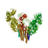
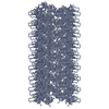
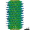
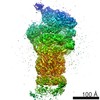
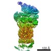
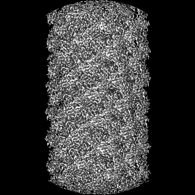
| Title | Structure of Salmonella flagellar hook reveals intermolecular domain interactions for the universal joint function | |||||||||
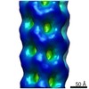
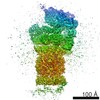
 Map data Map data | ||||||||||
 Sample Sample |
| |||||||||
 Keywords Keywords | FlgE / universal joint / axial structure / motor protein | |||||||||
| Function / homology |  Function and homology information Function and homology informationbacterial-type flagellum hook / bacterial-type flagellum basal body / bacterial-type flagellum-dependent swarming motility / bacterial-type flagellum / cytosol Similarity search - Function | |||||||||
| Biological species |  Salmonella enterica subsp. enterica serovar Typhimurium (bacteria) Salmonella enterica subsp. enterica serovar Typhimurium (bacteria) | |||||||||
| Method | helical reconstruction / cryo EM / Resolution: 4.1 Å | |||||||||
 Authors Authors | Horvath P / Kato T | |||||||||
| Funding support |  Japan, 2 items Japan, 2 items
| |||||||||
 Citation Citation | Journal: Biomolecules / Year: 2019 Title: Structure of Flagellar Hook Reveals Intermolecular Domain Interactions for the Universal Joint Function. Authors: Péter Horváth / Takayuki Kato / Tomoko Miyata / Keiichi Namba / Abstract: The bacterial flagellum is a motility organelle consisting of a rotary motor and a long helical filament as a propeller. The flagellar hook is a flexible universal joint that transmits motor torque ...The bacterial flagellum is a motility organelle consisting of a rotary motor and a long helical filament as a propeller. The flagellar hook is a flexible universal joint that transmits motor torque to the filament in its various orientations that change dynamically between swimming and tumbling of the cell upon switching the motor rotation for chemotaxis. Although the structures of the hook and hook protein FlgE from different bacterial species have been studied, the structure of hook, which has been studied most over the years, has not been solved at a high enough resolution to allow building an atomic model of entire FlgE for understanding the mechanisms of self-assembly, stability and the universal joint function. Here we report the structure of polyhook at 4.1 Å resolution by electron cryomicroscopy and helical image analysis. The density map clearly revealed folding of the entire FlgE chain forming the three domains D0, D1 and D2 and allowed us to build an atomic model. The model includes domain Dc with a long β-hairpin structure that connects domains D0 and D1 and contributes to the structural stability of the hook while allowing the flexible bending of the hook as a molecular universal joint. | |||||||||
| History |
|
- Structure visualization
| Movie |
Movie viewer |
|---|---|
| Structure viewer | EM map: SurfViewMolmilJmol/JSmol |
| Supplemental images |

- Downloads & links
-EMDB archive
| Map data | emd_9974.map.gz | 210.6 MB | EMDB map data format | |
|---|---|---|---|---|
| Header (meta data) | emd-9974-v30.xmlemd-9974.xml | 13.4 KB 13.4 KB | Display Display | EMDB header |
| Images | 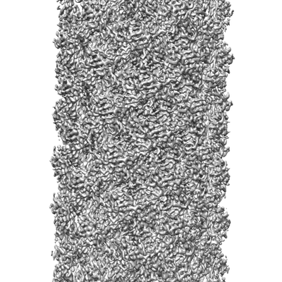 emd_9974.png emd_9974.png | 95.9 KB | ||
| Filedesc metadata | emd-9974.cif.gz | 5.7 KB | ||
| Archive directory |  http://ftp.pdbj.org/pub/emdb/structures/EMD-9974ftp://ftp.pdbj.org/pub/emdb/structures/EMD-9974 http://ftp.pdbj.org/pub/emdb/structures/EMD-9974ftp://ftp.pdbj.org/pub/emdb/structures/EMD-9974 | HTTPS FTP |
-Related structure data
| Related structure data |  6kfkMC M: atomic model generated by this map C: citing same article ( |
|---|---|
| Similar structure data |
-Links
| EMDB pages | EMDB (EBI/PDBe) / EMDataResource |
|---|---|
| Related items in Molecule of the Month |

-Map
| File | Download / File: emd_9974.map.gz / Format: CCP4 / Size: 229.8 MB / Type: IMAGE STORED AS FLOATING POINT NUMBER (4 BYTES) | ||||||||||||||||||||||||||||||||||||||||||||||||||||||||||||
|---|---|---|---|---|---|---|---|---|---|---|---|---|---|---|---|---|---|---|---|---|---|---|---|---|---|---|---|---|---|---|---|---|---|---|---|---|---|---|---|---|---|---|---|---|---|---|---|---|---|---|---|---|---|---|---|---|---|---|---|---|---|
| Projections & slices | Image control
Images are generated by Spider. | ||||||||||||||||||||||||||||||||||||||||||||||||||||||||||||
| Voxel size | X=Y=Z: 0.82 Å | ||||||||||||||||||||||||||||||||||||||||||||||||||||||||||||
| Density |
| ||||||||||||||||||||||||||||||||||||||||||||||||||||||||||||
| Symmetry | Space group: 1 | ||||||||||||||||||||||||||||||||||||||||||||||||||||||||||||
| Details | EMDB XML:
CCP4 map header:
| ||||||||||||||||||||||||||||||||||||||||||||||||||||||||||||
 Z (Sec.)
 Y (Row.)
 X (Col.)


-Supplemental data
- Sample components
-Entire : Flagellar hook
| Entire | Name: Flagellar hook |
|---|---|
| Components |
|
-Supramolecule #1: Flagellar hook
| Supramolecule | Name: Flagellar hook / type: complex / ID: 1 / Parent: 0 / Macromolecule list: all |
|---|---|
| Source (natural) | Organism: Salmonella enterica subsp. enterica serovar Typhimurium (bacteria) |
| Molecular weight | Theoretical: 42 kDa/nm |
-Macromolecule #1: Flagellar hook protein FlgE
| Macromolecule | Name: Flagellar hook protein FlgE / type: protein_or_peptide / ID: 1 / Number of copies: 1 / Enantiomer: LEVO |
|---|---|
| Source (natural) | Organism: Salmonella enterica subsp. enterica serovar Typhimurium (bacteria) |
| Molecular weight | Theoretical: 42.101957 KDa |
| Sequence | String: SFSQAVSGLN AAATNLDVIG NNIANSATYG FKSGTASFAD MFAGSKVGLG VKVAGITQDF TDGTTTNTGR GLDVAISQNG FFRLVDSNG SVFYSRNGQF KLDENRNLVN MQGMQLTGYP ATGTPPTIQQ GANPAPITIP NTLMAAKSTT TASMQINLNS T DPVPSKTP ...String: SFSQAVSGLN AAATNLDVIG NNIANSATYG FKSGTASFAD MFAGSKVGLG VKVAGITQDF TDGTTTNTGR GLDVAISQNG FFRLVDSNG SVFYSRNGQF KLDENRNLVN MQGMQLTGYP ATGTPPTIQQ GANPAPITIP NTLMAAKSTT TASMQINLNS T DPVPSKTP FSVSDADSYN KKGTVTVYDS QGNAHDMNVY FVKTKDNEWA VYTHDSSDPA ATAPTTASTT LKFNENGILE SG GTVNITT GTINGATAAT FSLSFLNSMQ QNTGANNIVA TNQNGYKPGD LVSYQINNDG TVVGNYSNEQ EQVLGQIVLA NFA NNEGLA SQGDNVWAAT QASGVALLGT AGSGNFGKLT NGALEASNVD LSKELVNMIV AQRNYQSNAQ TIKTQDQILN TLVN LR UniProtKB: Flagellar hook protein FlgE |
-Experimental details
-Structure determination
| Method | cryo EM |
|---|---|
 Processing Processing | helical reconstruction |
| Aggregation state | filament |
-Sample preparation
| Concentration | 0.8 mg/mL | |||||||||
|---|---|---|---|---|---|---|---|---|---|---|
| Buffer | pH: 8 Component:
| |||||||||
| Grid | Model: Quantifoil R0.6/1 / Material: MOLYBDENUM / Mesh: 200 / Pretreatment - Type: GLOW DISCHARGE / Pretreatment - Time: 30 sec. | |||||||||
| Vitrification | Cryogen name: ETHANE / Chamber humidity: 100 % / Chamber temperature: 275.15 K |
- Electron microscopy
| Microscope | JEOL 3200FSC |
|---|---|
| Specialist optics | Energy filter - Name: In-column Omega Filter / Energy filter - Slit width: 10 eV |
| Image recording | Film or detector model: GATAN K2 SUMMIT (4k x 4k) / Detector mode: SUPER-RESOLUTION / Digitization - Dimensions - Width: 7676 pixel / Digitization - Dimensions - Height: 7420 pixel / Number real images: 486 / Average exposure time: 6.0 sec. / Average electron dose: 30.0 e/Å2 |
| Electron beam | Acceleration voltage: 300 kV / Electron source:  FIELD EMISSION GUN FIELD EMISSION GUN |
| Electron optics | Illumination mode: FLOOD BEAM / Imaging mode: BRIGHT FIELD / Cs: 4.1 mm / Nominal magnification: 50000 |
| Sample stage | Cooling holder cryogen: NITROGEN |
-Image processing
| Final reconstruction | Applied symmetry - Helical parameters - Δz: 4.05 Å Applied symmetry - Helical parameters - Δ&Phi: 64.78 ° Applied symmetry - Helical parameters - Axial symmetry: C1 (asymmetric) Resolution.type: BY AUTHOR / Resolution: 4.1 Å / Resolution method: FSC 0.143 CUT-OFF / Software - Name: RELION (ver. 2) / Number images used: 41568 |
|---|---|
| Segment selection | Number selected: 42293 |
| Startup model | Type of model: OTHER |
| Final angle assignment | Type: NOT APPLICABLE / Software - Name: RELION (ver. 2) |
-Atomic model buiding 1
| Initial model |
| ||||||||
|---|---|---|---|---|---|---|---|---|---|
| Refinement | Space: RECIPROCAL / Protocol: FLEXIBLE FIT | ||||||||
| Output model | PDB-6kfk: |