 Movie
Movie Controller


+ Open data

- Basic information
| Entry | Database: EMDB / ID: EMD-9911 | |||||||||
|---|---|---|---|---|---|---|---|---|---|---|
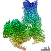
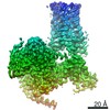
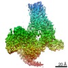
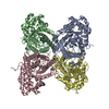
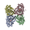
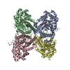
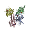
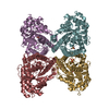
| Title | cryo-EM structure of alpha2BAR-GoA complex | |||||||||
 Map data Map data | alpha2BAR-GoA complex | |||||||||
 Sample Sample |
| |||||||||
 Keywords Keywords | GPCR / Complex / cryo-EM / MEMBRANE PROTEIN | |||||||||
| Function / homology |  Function and homology information Function and homology informationAdrenoceptors / Adrenaline signalling through Alpha-2 adrenergic receptor / Surfactant metabolism / Activation of the phototransduction cascade / Olfactory Signaling Pathway / G beta:gamma signalling through PLC beta / Presynaptic function of Kainate receptors / adenylate cyclase-inhibiting adrenergic receptor signaling pathway / Prostacyclin signalling through prostacyclin receptor / Sensory perception of sweet, bitter, and umami (glutamate) taste ...Adrenoceptors / Adrenaline signalling through Alpha-2 adrenergic receptor / Surfactant metabolism / Activation of the phototransduction cascade / Olfactory Signaling Pathway / G beta:gamma signalling through PLC beta / Presynaptic function of Kainate receptors / adenylate cyclase-inhibiting adrenergic receptor signaling pathway / Prostacyclin signalling through prostacyclin receptor / Sensory perception of sweet, bitter, and umami (glutamate) taste / alpha2-adrenergic receptor activity / Adrenaline signalling through Alpha-2 adrenergic receptor / Synthesis, secretion, and inactivation of Glucagon-like Peptide-1 (GLP-1) / G alpha (z) signalling events / Glucagon-type ligand receptors / G beta:gamma signalling through PI3Kgamma / G beta:gamma signalling through CDC42 / alpha-2C adrenergic receptor binding / epinephrine binding / positive regulation of uterine smooth muscle contraction / Adrenaline,noradrenaline inhibits insulin secretion / phospholipase C-activating adrenergic receptor signaling pathway / ADP signalling through P2Y purinoceptor 12 / Cooperation of PDCL (PhLP1) and TRiC/CCT in G-protein beta folding / G beta:gamma signalling through BTK / Thromboxane signalling through TP receptor / Thrombin signalling through proteinase activated receptors (PARs) / Activation of G protein gated Potassium channels / Inhibition of voltage gated Ca2+ channels via Gbeta/gamma subunits / negative regulation of norepinephrine secretion / G-protein activation / G alpha (s) signalling events / negative regulation of epinephrine secretion / Ca2+ pathway / G alpha (12/13) signalling events / Extra-nuclear estrogen signaling / G alpha (q) signalling events / Vasopressin regulates renal water homeostasis via Aquaporins / GPER1 signaling / regulation of vascular associated smooth muscle contraction / Glucagon-like Peptide-1 (GLP1) regulates insulin secretion / heterotrimeric G-protein binding / G alpha (i) signalling events / High laminar flow shear stress activates signaling by PIEZO1 and PECAM1:CDH5:KDR in endothelial cells / ADP signalling through P2Y purinoceptor 1 / positive regulation of blood pressure / regulation of smooth muscle contraction / mu-type opioid receptor binding / corticotropin-releasing hormone receptor 1 binding / dopaminergic synapse / fear response / thioesterase binding / negative regulation of insulin secretion involved in cellular response to glucose stimulus / vesicle docking involved in exocytosis / positive regulation of membrane protein ectodomain proteolysis / norepinephrine binding / Adrenoceptors / Adrenaline,noradrenaline inhibits insulin secretion / G protein-coupled dopamine receptor signaling pathway / positive regulation of epidermal growth factor receptor signaling pathway / G alpha (z) signalling events / positive regulation of wound healing / spectrin binding / adrenergic receptor signaling pathway / G alpha (i) signalling events / regulation of heart contraction / parallel fiber to Purkinje cell synapse / alkylglycerophosphoethanolamine phosphodiesterase activity / phototransduction, visible light / photoreceptor outer segment / regulation of vasoconstriction / postsynaptic modulation of chemical synaptic transmission / negative regulation of lipid catabolic process / viral release from host cell by cytolysis / cellular response to hormone stimulus / adenylate cyclase-activating adrenergic receptor signaling pathway / adenylate cyclase regulator activity / G protein-coupled serotonin receptor binding / adenylate cyclase-inhibiting serotonin receptor signaling pathway / peptidoglycan catabolic process / cardiac muscle cell apoptotic process / positive regulation of neuron differentiation / presynaptic modulation of chemical synaptic transmission / muscle contraction / positive regulation of cytokine production / locomotory behavior / female pregnancy / negative regulation of insulin secretion / GABA-ergic synapse / platelet activation / adenylate cyclase-modulating G protein-coupled receptor signaling pathway / G-protein beta/gamma-subunit complex binding / G beta:gamma signalling through PLC beta / Presynaptic function of Kainate receptors / Thromboxane signalling through TP receptor / Activation of G protein gated Potassium channels / Inhibition of voltage gated Ca2+ channels via Gbeta/gamma subunits / G-protein activation / Prostacyclin signalling through prostacyclin receptor / G beta:gamma signalling through CDC42 Similarity search - Function | |||||||||
| Biological species |  Spodoptera (butterflies/moths) / Spodoptera (butterflies/moths) /  Homo sapiens (human) / Homo sapiens (human) /  | |||||||||
| Method | single particle reconstruction / cryo EM / Resolution: 2.9 Å | |||||||||
 Authors Authors | Yuan D / Liu Z | |||||||||
 Citation Citation | Journal: Nat Chem Biol / Year: 2020 Title: Activation of the α adrenoceptor by the sedative sympatholytic dexmedetomidine. Authors: Daopeng Yuan / Zhongmin Liu / Jonas Kaindl / Shoji Maeda / Jiawei Zhao / Xiaoou Sun / Jun Xu / Peter Gmeiner / Hong-Wei Wang / Brian K Kobilka /    Abstract: The α adrenergic receptors (αARs) are G protein-coupled receptors (GPCRs) that respond to adrenaline and noradrenaline and couple to the Gi/o family of G proteins. αARs play important roles in ...The α adrenergic receptors (αARs) are G protein-coupled receptors (GPCRs) that respond to adrenaline and noradrenaline and couple to the Gi/o family of G proteins. αARs play important roles in regulating the sympathetic nervous system. Dexmedetomidine is a highly selective αAR agonist used in post-operative patients as an anxiety-reducing, sedative medicine that decreases the requirement for opioids. As is typical for selective αAR agonists, dexmedetomidine consists of an imidazole ring and a substituted benzene moiety lacking polar groups, which is in contrast to βAR-selective agonists, which share an ethanolamine group and an aromatic system with polar, hydrogen-bonding substituents. To better understand the structural basis for the selectivity and efficacy of adrenergic agonists, we determined the structure of the αAR in complex with dexmedetomidine and Go at a resolution of 2.9 Å by single-particle cryo-EM. The structure reveals the mechanism of αAR-selective activation and provides insights into Gi/o coupling specificity. | |||||||||
| History |
|
- Structure visualization
| Movie |
Movie viewer |
|---|---|
| Structure viewer | EM map: SurfViewMolmilJmol/JSmol |
| Supplemental images |

- Downloads & links
-EMDB archive
| Map data | emd_9911.map.gz | 15.8 MB | EMDB map data format | |
|---|---|---|---|---|
| Header (meta data) | emd-9911-v30.xmlemd-9911.xml | 19.1 KB 19.1 KB | Display Display | EMDB header |
| Images | 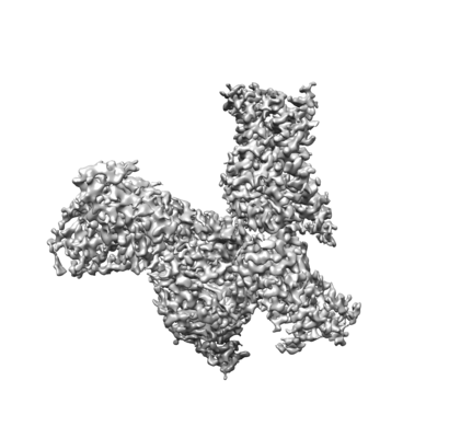 emd_9911.png emd_9911.png | 54.1 KB | ||
| Filedesc metadata | emd-9911.cif.gz | 7.1 KB | ||
| Archive directory |  http://ftp.pdbj.org/pub/emdb/structures/EMD-9911ftp://ftp.pdbj.org/pub/emdb/structures/EMD-9911 http://ftp.pdbj.org/pub/emdb/structures/EMD-9911ftp://ftp.pdbj.org/pub/emdb/structures/EMD-9911 | HTTPS FTP |
-Validation report
| Summary document | emd_9911_validation.pdf.gz | 363.6 KB | Display | EMDB validaton report |
|---|---|---|---|---|
| Full document | emd_9911_full_validation.pdf.gz | 363.2 KB | Display | |
| Data in XML | emd_9911_validation.xml.gz | 7.1 KB | Display | |
| Data in CIF | emd_9911_validation.cif.gz | 8.2 KB | Display | |
| Arichive directory | https://ftp.pdbj.org/pub/emdb/validation_reports/EMD-9911ftp://ftp.pdbj.org/pub/emdb/validation_reports/EMD-9911 | HTTPS FTP |
-Related structure data
| Related structure data | 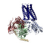 6k41MC 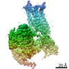 9912C 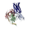 6k42C C: citing same article ( M: atomic model generated by this map |
|---|---|
| Similar structure data |

-Links
| EMDB pages | EMDB (EBI/PDBe) / EMDataResource |
|---|---|
| Related items in Molecule of the Month |


-Map
| File | Download / File: emd_9911.map.gz / Format: CCP4 / Size: 244.1 MB / Type: IMAGE STORED AS FLOATING POINT NUMBER (4 BYTES) | ||||||||||||||||||||||||||||||||||||||||||||||||||||||||||||||||||||
|---|---|---|---|---|---|---|---|---|---|---|---|---|---|---|---|---|---|---|---|---|---|---|---|---|---|---|---|---|---|---|---|---|---|---|---|---|---|---|---|---|---|---|---|---|---|---|---|---|---|---|---|---|---|---|---|---|---|---|---|---|---|---|---|---|---|---|---|---|---|
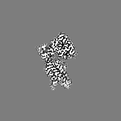
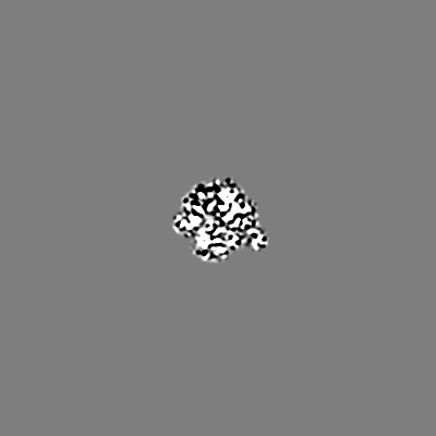
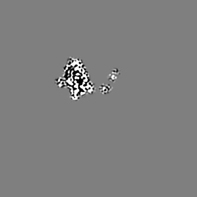
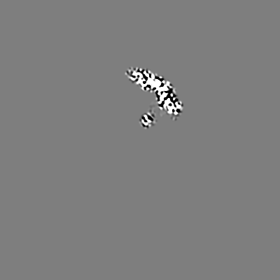
| Annotation | alpha2BAR-GoA complex | ||||||||||||||||||||||||||||||||||||||||||||||||||||||||||||||||||||
| Projections & slices | Image control
Images are generated by Spider. | ||||||||||||||||||||||||||||||||||||||||||||||||||||||||||||||||||||
| Voxel size | X=Y=Z: 0.5455 Å | ||||||||||||||||||||||||||||||||||||||||||||||||||||||||||||||||||||
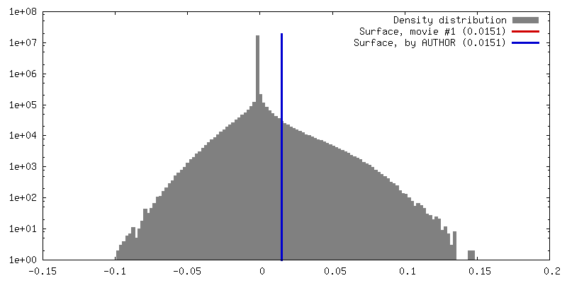
| Density |
| ||||||||||||||||||||||||||||||||||||||||||||||||||||||||||||||||||||
| Symmetry | Space group: 1 | ||||||||||||||||||||||||||||||||||||||||||||||||||||||||||||||||||||
| Details | EMDB XML:
CCP4 map header:
| ||||||||||||||||||||||||||||||||||||||||||||||||||||||||||||||||||||
 Z (Sec.)
 Y (Row.)
 X (Col.)


-Supplemental data
- Sample components
-Entire : alpha2BAR-GoA complex
| Entire | Name: alpha2BAR-GoA complex |
|---|---|
| Components |
|
-Supramolecule #1: alpha2BAR-GoA complex
| Supramolecule | Name: alpha2BAR-GoA complex / type: complex / ID: 1 / Parent: 0 / Macromolecule list: #1-#5 |
|---|---|
| Source (natural) | Organism: Spodoptera (butterflies/moths) |
| Molecular weight | Theoretical: 150 KDa |
-Macromolecule #1: Guanine nucleotide-binding protein G(o) subunit alpha
| Macromolecule | Name: Guanine nucleotide-binding protein G(o) subunit alpha / type: protein_or_peptide / ID: 1 / Number of copies: 1 / Enantiomer: LEVO |
|---|---|
| Source (natural) | Organism: Homo sapiens (human) |
| Molecular weight | Theoretical: 40.1005 KDa |
| Recombinant expression | Organism: Spodoptera (butterflies/moths) |
| Sequence | String: MGCTLSAEER AALERSKAIE KNLKEDGISA AKDVKLLLLG AGESGKSTIV KQMKIIHEDG FSGEDVKQYK PVVYSNTIQS LAAIVRAMD TLGIEYGDKE RKADAKMVCD VVSRMEDTEP FSAELLSAMM RLWGDSGIQE CFNRSREYQL NDSAKYYLDS L DRIGAADY ...String: MGCTLSAEER AALERSKAIE KNLKEDGISA AKDVKLLLLG AGESGKSTIV KQMKIIHEDG FSGEDVKQYK PVVYSNTIQS LAAIVRAMD TLGIEYGDKE RKADAKMVCD VVSRMEDTEP FSAELLSAMM RLWGDSGIQE CFNRSREYQL NDSAKYYLDS L DRIGAADY QPTEQDILRT RVKTTGIVET HFTFKNLHFR LFDVGGQRSE RKKWIHCFED VTAIIFCVAL SGYDQVLHED ET TNRMHES LMLFDSICNN KFFIDTSIIL FLNKKDLFGE KIKKSPLTIC FPEYTGPNTY EDAAAYIQAQ FESKNRSPNK EIY CHMTCA TDTNNIQVVF DAVTDIIIAN NLRGCGLY UniProtKB: Guanine nucleotide-binding protein G(o) subunit alpha |
-Macromolecule #2: Guanine nucleotide-binding protein G(I)/G(S)/G(T) subunit beta-1
| Macromolecule | Name: Guanine nucleotide-binding protein G(I)/G(S)/G(T) subunit beta-1 type: protein_or_peptide / ID: 2 / Number of copies: 1 / Enantiomer: LEVO |
|---|---|
| Source (natural) | Organism: |
| Molecular weight | Theoretical: 38.402867 KDa |
| Recombinant expression | Organism: Spodoptera (butterflies/moths) |
| Sequence | String: HHHHHHGSSG SELDQLRQEA EQLKNQIRDA RKACADATLS QITNNIDPVG RIQMRTRRTL RGHLAKIYAM HWGTDSRLLV SASQDGKLI IWDSYTTNKV HAIPLRSSWV MTCAYAPSGN YVACGGLDNI CSIYNLKTRE GNVRVSRELA GHTGYLSCCR F LDDNQIVT ...String: HHHHHHGSSG SELDQLRQEA EQLKNQIRDA RKACADATLS QITNNIDPVG RIQMRTRRTL RGHLAKIYAM HWGTDSRLLV SASQDGKLI IWDSYTTNKV HAIPLRSSWV MTCAYAPSGN YVACGGLDNI CSIYNLKTRE GNVRVSRELA GHTGYLSCCR F LDDNQIVT SSGDTTCALW DIETGQQTTT FTGHTGDVMS LSLAPDTRLF VSGACDASAK LWDVREGMCR QTFTGHESDI NA ICFFPNG NAFATGSDDA TCRLFDLRAD QELMTYSHDN IICGITSVSF SKSGRLLLAG YDDFNCNVWD ALKADRAGVL AGH DNRVSC LGVTDDGMAV ATGSWDSFLK IWN UniProtKB: Guanine nucleotide-binding protein G(I)/G(S)/G(T) subunit beta-1 |
-Macromolecule #3: Guanine nucleotide-binding protein G(I)/G(S)/G(O) subunit gamma-2
| Macromolecule | Name: Guanine nucleotide-binding protein G(I)/G(S)/G(O) subunit gamma-2 type: protein_or_peptide / ID: 3 / Number of copies: 1 / Enantiomer: LEVO |
|---|---|
| Source (natural) | Organism: Homo sapiens (human) |
| Molecular weight | Theoretical: 7.861143 KDa |
| Recombinant expression | Organism: Spodoptera (butterflies/moths) |
| Sequence | String: MASNNTASIA QARKLVEQLK MEANIDRIKV SKAAADLMAY CEAHAKEDPL LTPVPASENP FREKKFFCAI L UniProtKB: Guanine nucleotide-binding protein G(I)/G(S)/G(O) subunit gamma-2 |
-Macromolecule #4: Alpha-2A adrenergic receptor,Endolysin,Alpha-2B adrenergic recept...
| Macromolecule | Name: Alpha-2A adrenergic receptor,Endolysin,Alpha-2B adrenergic receptor,Alpha-2B adrenergic receptor type: protein_or_peptide / ID: 4 / Number of copies: 1 / Enantiomer: LEVO / EC number: lysozyme |
|---|---|
| Source (natural) | Organism: Homo sapiens (human) |
| Molecular weight | Theoretical: 58.156211 KDa |
| Recombinant expression | Organism: Spodoptera (butterflies/moths) |
| Sequence | String: DDDDAHHHHH HMGSLQPDAG NASWNGTEAP GGGARATPEN LYFQGNIFEM LRIDEGLRLK IYKDTEGYYT IGIGHLLTKS PSLNAAKSE LDKAIGRNTN GVITKDEAEK LFNQDVDAAV RGILRNAKLK PVYDSLDAVR RAALINMVFQ MGETGVAGFT N SLRMLQQK ...String: DDDDAHHHHH HMGSLQPDAG NASWNGTEAP GGGARATPEN LYFQGNIFEM LRIDEGLRLK IYKDTEGYYT IGIGHLLTKS PSLNAAKSE LDKAIGRNTN GVITKDEAEK LFNQDVDAAV RGILRNAKLK PVYDSLDAVR RAALINMVFQ MGETGVAGFT N SLRMLQQK RWDEAAVNLA KSRWYNQTPN RAKRVITTFR TGTWDAYYSV QATAAIAAAI TFLILFTIFG NALVILAVLT SR SLRAPQN LFLVSLAAAD ILVATLIIPF SLANELLGYW YFRRTWCEVY LALDVLFCTS SIVHLCAISL DRYWAVSRAL EYN SKRTPR RIKCIILTVW LIAAVISLPP LIYKGDQGPQ PRGRPQCKLN QEAWYILASS IGSFFAPCLI MILVYLRIYL IAKR SNRRG PRAKGGPGQG EQWWRRRAQL TREKRFTFVL AVVIGVFVLC WFPFFFSYSL GAICPKHCKV PHGLFQFFFW IGYCN SSLN PVIYTIFNQD FRRAFRRILC RPWTQTAW UniProtKB: Alpha-2A adrenergic receptor, Endolysin, Alpha-2B adrenergic receptor, Alpha-2B adrenergic receptor |
-Macromolecule #5: scFv
| Macromolecule | Name: scFv / type: protein_or_peptide / ID: 5 / Number of copies: 1 / Enantiomer: LEVO |
|---|---|
| Source (natural) | Organism: |
| Molecular weight | Theoretical: 32.898781 KDa |
| Recombinant expression | Organism: Spodoptera (butterflies/moths) |
| Sequence | String: MLLVNQSHQG FNKEHTSKMV SAIVLYVLLA AAAHSAFADV QLVESGGGLV QPGGSRKLSC SASGFAFSSF GMHWVRQAPE KGLEWVAYI SSGSGTIYYA DTVKGRFTIS RDDPKNTLFL QMTSLRSEDT AMYYCVRSIY YYGSSPFDFW GQGTTLTVSS G GGGSGGGG ...String: MLLVNQSHQG FNKEHTSKMV SAIVLYVLLA AAAHSAFADV QLVESGGGLV QPGGSRKLSC SASGFAFSSF GMHWVRQAPE KGLEWVAYI SSGSGTIYYA DTVKGRFTIS RDDPKNTLFL QMTSLRSEDT AMYYCVRSIY YYGSSPFDFW GQGTTLTVSS G GGGSGGGG SGGGGSDIVM TQATSSVPVT PGESVSISCR SSKSLLHSNG NTYLYWFLQR PGQSPQLLIY RMSNLASGVP DR FSGSGSG TAFTLTISRL EAEDVGVYYC MQHLEYPLTF GAGTKLELKG SLEVLFQGPA AAHHHHHHHH |
-Macromolecule #6: 4-[(1~{S})-1-(2,3-dimethylphenyl)ethyl]-1~{H}-imidazole
| Macromolecule | Name: 4-[(1~{S})-1-(2,3-dimethylphenyl)ethyl]-1~{H}-imidazole type: ligand / ID: 6 / Number of copies: 1 / Formula: CZX |
|---|---|
| Molecular weight | Theoretical: 200.28 Da |
| Chemical component information |  ChemComp-CZX: |
-Experimental details
-Structure determination
| Method | cryo EM |
|---|---|
 Processing Processing | single particle reconstruction |
| Aggregation state | particle |
-Sample preparation
| Concentration | 10 mg/mL |
|---|---|
| Buffer | pH: 7.5 |
| Grid | Model: Quantifoil R1.2/1.3 / Material: GOLD / Mesh: 400 |
| Vitrification | Cryogen name: ETHANE / Chamber humidity: 100 % / Instrument: FEI VITROBOT MARK IV |
- Electron microscopy
| Microscope | FEI TITAN KRIOS |
|---|---|
| Image recording | Film or detector model: GATAN K2 SUMMIT (4k x 4k) / Detector mode: SUPER-RESOLUTION / Average electron dose: 50.0 e/Å2 |
| Electron beam | Acceleration voltage: 300 kV / Electron source:  FIELD EMISSION GUN FIELD EMISSION GUN |
| Electron optics | Illumination mode: SPOT SCAN / Imaging mode: BRIGHT FIELD |
| Experimental equipment |  Model: Titan Krios / Image courtesy: FEI Company |
