+ データを開く
- 基本情報
基本情報
| 登録情報 | データベース: EMDB / ID: EMD-9572 | |||||||||
|---|---|---|---|---|---|---|---|---|---|---|
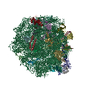
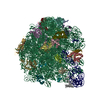
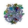
| タイトル | Structure of the large subunit of the chloro-ribosome | |||||||||
 マップデータ マップデータ | Chloro-ribosome LSU (50S) | |||||||||
 試料 試料 |
| |||||||||
 キーワード キーワード | Cryo-EM / ribosome / chloro-ribosome | |||||||||
| 機能・相同性 |  機能・相同性情報 機能・相同性情報plastid translation / chloroplast envelope / mitochondrial large ribosomal subunit / mitochondrial translation / chloroplast thylakoid membrane / chloroplast / DNA-templated transcription termination / large ribosomal subunit / transferase activity / 5S rRNA binding ...plastid translation / chloroplast envelope / mitochondrial large ribosomal subunit / mitochondrial translation / chloroplast thylakoid membrane / chloroplast / DNA-templated transcription termination / large ribosomal subunit / transferase activity / 5S rRNA binding / ribosomal large subunit assembly / large ribosomal subunit rRNA binding / cytosolic large ribosomal subunit / negative regulation of translation / rRNA binding / structural constituent of ribosome / ribosome / translation / ribonucleoprotein complex / mRNA binding / mitochondrion / RNA binding 類似検索 - 分子機能 | |||||||||
| 生物種 |  Spinacia oleracea (ホウレンソウ) Spinacia oleracea (ホウレンソウ) | |||||||||
| 手法 | 単粒子再構成法 / クライオ電子顕微鏡法 / 解像度: 3.5 Å | |||||||||
 データ登録者 データ登録者 | Ahmed T / Yin Z | |||||||||
| 資金援助 |  シンガポール, 1件 シンガポール, 1件
| |||||||||
 引用 引用 |  ジャーナル: Sci Rep / 年: 2016 ジャーナル: Sci Rep / 年: 2016タイトル: Cryo-EM structure of the large subunit of the spinach chloroplast ribosome. 著者: Tofayel Ahmed / Zhan Yin / Shashi Bhushan /  要旨: Protein synthesis in the chloroplast is mediated by the chloroplast ribosome (chloro-ribosome). Overall architecture of the chloro-ribosome is considerably similar to the Escherichia coli (E. coli) ...Protein synthesis in the chloroplast is mediated by the chloroplast ribosome (chloro-ribosome). Overall architecture of the chloro-ribosome is considerably similar to the Escherichia coli (E. coli) ribosome but certain differences are evident. The chloro-ribosome proteins are generally larger because of the presence of chloroplast-specific extensions in their N- and C-termini. The chloro-ribosome harbours six plastid-specific ribosomal proteins (PSRPs); four in the small subunit and two in the large subunit. Deletions and insertions occur throughout the rRNA sequence of the chloro-ribosome (except for the conserved peptidyl transferase center region) but the overall length of the rRNAs do not change significantly, compared to the E. coli. Although, recent advancements in cryo-electron microscopy (cryo-EM) have provided detailed high-resolution structures of ribosomes from many different sources, a high-resolution structure of the chloro-ribosome is still lacking. Here, we present a cryo-EM structure of the large subunit of the chloro-ribosome from spinach (Spinacia oleracea) at an average resolution of 3.5 Å. High-resolution map enabled us to localize and model chloro-ribosome proteins, chloroplast-specific protein extensions, two PSRPs (PSRP5 and 6) and three rRNA molecules present in the chloro-ribosome. Although comparable to E. coli, the polypeptide tunnel and the tunnel exit site show chloroplast-specific features. | |||||||||
| 履歴 |
|
- 構造の表示
構造の表示
| ムービー |
 ムービービューア ムービービューア |
|---|---|
| 構造ビューア | EMマップ:  SurfView SurfView Molmil Molmil Jmol/JSmol Jmol/JSmol |
| 添付画像 |
- ダウンロードとリンク
-EMDBアーカイブ
| マップデータ |  emd_9572.map.gz emd_9572.map.gz | 11.6 MB |  EMDBマップデータ形式 EMDBマップデータ形式 | |
|---|---|---|---|---|
| ヘッダ (付随情報) |  emd-9572-v30.xml emd-9572-v30.xml emd-9572.xml emd-9572.xml | 49.8 KB 49.8 KB | 表示 表示 |  EMDBヘッダ EMDBヘッダ |
| 画像 |  emd_9572.png emd_9572.png | 63.7 KB | ||
| Filedesc metadata |  emd-9572.cif.gz emd-9572.cif.gz | 11.3 KB | ||
| アーカイブディレクトリ |  http://ftp.pdbj.org/pub/emdb/structures/EMD-9572 http://ftp.pdbj.org/pub/emdb/structures/EMD-9572 ftp://ftp.pdbj.org/pub/emdb/structures/EMD-9572 ftp://ftp.pdbj.org/pub/emdb/structures/EMD-9572 | HTTPS FTP |
-検証レポート
| 文書・要旨 |  emd_9572_validation.pdf.gz emd_9572_validation.pdf.gz | 429.2 KB | 表示 |  EMDB検証レポート EMDB検証レポート |
|---|---|---|---|---|
| 文書・詳細版 |  emd_9572_full_validation.pdf.gz emd_9572_full_validation.pdf.gz | 428.8 KB | 表示 | |
| XML形式データ |  emd_9572_validation.xml.gz emd_9572_validation.xml.gz | 6.7 KB | 表示 | |
| CIF形式データ |  emd_9572_validation.cif.gz emd_9572_validation.cif.gz | 7.7 KB | 表示 | |
| アーカイブディレクトリ |  https://ftp.pdbj.org/pub/emdb/validation_reports/EMD-9572 https://ftp.pdbj.org/pub/emdb/validation_reports/EMD-9572 ftp://ftp.pdbj.org/pub/emdb/validation_reports/EMD-9572 ftp://ftp.pdbj.org/pub/emdb/validation_reports/EMD-9572 | HTTPS FTP |
-関連構造データ
- リンク
リンク
| EMDBのページ |  EMDB (EBI/PDBe) / EMDB (EBI/PDBe) /  EMDataResource EMDataResource |
|---|---|
| 「今月の分子」の関連する項目 |
- マップ
マップ
| ファイル |  ダウンロード / ファイル: emd_9572.map.gz / 形式: CCP4 / 大きさ: 115.9 MB / タイプ: IMAGE STORED AS FLOATING POINT NUMBER (4 BYTES) ダウンロード / ファイル: emd_9572.map.gz / 形式: CCP4 / 大きさ: 115.9 MB / タイプ: IMAGE STORED AS FLOATING POINT NUMBER (4 BYTES) | ||||||||||||||||||||||||||||||||||||||||||||||||||||||||||||
|---|---|---|---|---|---|---|---|---|---|---|---|---|---|---|---|---|---|---|---|---|---|---|---|---|---|---|---|---|---|---|---|---|---|---|---|---|---|---|---|---|---|---|---|---|---|---|---|---|---|---|---|---|---|---|---|---|---|---|---|---|---|
| 注釈 | Chloro-ribosome LSU (50S) | ||||||||||||||||||||||||||||||||||||||||||||||||||||||||||||
| 投影像・断面図 | 画像のコントロール
画像は Spider により作成 | ||||||||||||||||||||||||||||||||||||||||||||||||||||||||||||
| ボクセルのサイズ | X=Y=Z: 1.28 Å | ||||||||||||||||||||||||||||||||||||||||||||||||||||||||||||
| 密度 |
| ||||||||||||||||||||||||||||||||||||||||||||||||||||||||||||
| 対称性 | 空間群: 1 | ||||||||||||||||||||||||||||||||||||||||||||||||||||||||||||
| 詳細 | EMDB XML:
CCP4マップ ヘッダ情報:
| ||||||||||||||||||||||||||||||||||||||||||||||||||||||||||||
-添付データ
- 試料の構成要素
+全体 : Chloro-ribosome 50S
+超分子 #1: Chloro-ribosome 50S
+分子 #1: 23S rRNA
+分子 #2: Spinach chloroplast 4.5S rRNA
+分子 #3: 5S rRNA
+分子 #4: 50S ribosomal protein L13, chloroplastic
+分子 #5: 50S ribosomal protein L14, chloroplastic
+分子 #6: 50S ribosomal protein L15
+分子 #7: 50S ribosomal protein L16, chloroplastic
+分子 #8: 50S ribosomal protein L17
+分子 #9: 50S ribosomal protein L18
+分子 #10: 50S ribosomal protein L19, chloroplastic
+分子 #11: 50S ribosomal protein L20, chloroplastic
+分子 #12: 50S ribosomal protein L21, chloroplastic
+分子 #13: 50S ribosomal protein L22, chloroplastic
+分子 #14: 50S ribosomal protein L23, chloroplastic
+分子 #15: 50S ribosomal protein L24, chloroplastic
+分子 #16: 50S ribosomal protein L27
+分子 #17: 50S ribosomal protein L28
+分子 #18: 50S ribosomal protein L29
+分子 #19: 50S ribosomal protein L2, chloroplastic
+分子 #20: 50S ribosomal protein L32, chloroplastic
+分子 #21: 50S ribosomal protein L33, chloroplastic
+分子 #22: 50S ribosomal protein L34, chloroplastic
+分子 #23: 50S ribosomal protein L35, chloroplastic
+分子 #24: 50S ribosomal protein L36, chloroplastic
+分子 #25: 50S ribosomal protein L3
+分子 #26: 50S ribosomal protein L4, chloroplastic
+分子 #27: 50S ribosomal protein L5, chloroplastic
+分子 #28: 50S ribosomal protein L6
+分子 #29: 50S ribosomal protein L9
+分子 #30: 50S ribosomal protein 5 alpha, chloroplastic
+分子 #31: 50S ribosomal protein L31
+分子 #32: 50S ribosomal protein 6, chloroplastic
-実験情報
-構造解析
| 手法 | クライオ電子顕微鏡法 |
|---|---|
 解析 解析 | 単粒子再構成法 |
| 試料の集合状態 | particle |
- 試料調製
試料調製
| 緩衝液 | pH: 7.6 詳細: 20 mM Tris HCl, pH 7.6, 100 mM KCl, 10 mM MgOAc, 100 mM sucrose, 7 mM 2-mercaptoethanol, 1 unit/ml RNase inhibitor, 0.1% protease inhibitor |
|---|---|
| グリッド | モデル: Quantifoil / 材質: COPPER / メッシュ: 300 / 支持フィルム - 材質: CARBON / 支持フィルム - トポロジー: CONTINUOUS / 支持フィルム - Film thickness: 2 / 前処理 - タイプ: GLOW DISCHARGE / 前処理 - 時間: 30 sec. |
| 凍結 | 凍結剤: ETHANE / チャンバー内湿度: 100 % / チャンバー内温度: 277 K / 装置: FEI VITROBOT MARK IV |
- 電子顕微鏡法
| 顕微鏡 | FEI TECNAI ARCTICA |
|---|---|
| 撮影 | フィルム・検出器のモデル: FEI FALCON II (4k x 4k) 検出モード: INTEGRATING / デジタル化 - サイズ - 横: 4096 pixel / デジタル化 - サイズ - 縦: 4096 pixel / デジタル化 - 画像ごとのフレーム数: 1-7 / 撮影したグリッド数: 1 / 実像数: 1590 / 平均電子線量: 26.0 e/Å2 |
| 電子線 | 加速電圧: 200 kV / 電子線源:  FIELD EMISSION GUN FIELD EMISSION GUN |
| 電子光学系 | C2レンズ絞り径: 100.0 µm / 最大 デフォーカス(補正後): 2.5 µm / 最小 デフォーカス(補正後): 0.2 µm / 倍率(補正後): 109375 / 照射モード: FLOOD BEAM / 撮影モード: BRIGHT FIELD / Cs: 2.7 mm / 最大 デフォーカス(公称値): 2.5 µm / 最小 デフォーカス(公称値): 0.2 µm / 倍率(公称値): 78000 |
| 試料ステージ | 試料ホルダーモデル: FEI TITAN KRIOS AUTOGRID HOLDER ホルダー冷却材: NITROGEN |
| 実験機器 |  モデル: Talos Arctica / 画像提供: FEI Company |
 ムービー
ムービー コントローラー


 Z (Sec.)
 Y (Row.)
 X (Col.)


