 ムービー
ムービー コントローラー


+ データを開く

- 基本情報
基本情報
| 登録情報 | データベース: EMDB / ID: EMD-9217 | ||||||||||||
|---|---|---|---|---|---|---|---|---|---|---|---|---|---|
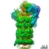
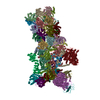
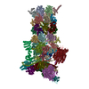
| タイトル | Cryo-EM structures and dynamics of substrate-engaged human 26S proteasome | ||||||||||||
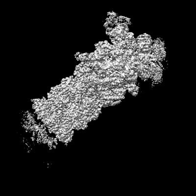
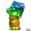
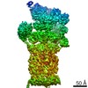
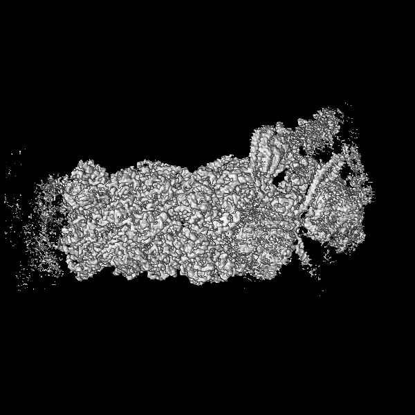
 マップデータ マップデータ | The complete map of state EA2 of substrate-engaged human proteasome, low pass-filtered to 3 Angstrom without amplitude correction. | ||||||||||||
 試料 試料 |
| ||||||||||||
 キーワード キーワード | Proteosome / HYDROLASE | ||||||||||||
| 機能・相同性 |  機能・相同性情報 機能・相同性情報thyrotropin-releasing hormone receptor binding / Impaired BRCA2 translocation to the nucleus / Impaired BRCA2 binding to SEM1 (DSS1) / nuclear proteasome complex / host-mediated perturbation of viral transcription / positive regulation of inclusion body assembly / integrator complex / proteasome accessory complex / meiosis I / purine ribonucleoside triphosphate binding ...thyrotropin-releasing hormone receptor binding / Impaired BRCA2 translocation to the nucleus / Impaired BRCA2 binding to SEM1 (DSS1) / nuclear proteasome complex / host-mediated perturbation of viral transcription / positive regulation of inclusion body assembly / integrator complex / proteasome accessory complex / meiosis I / purine ribonucleoside triphosphate binding / proteasome regulatory particle / cytosolic proteasome complex / hypothalamus gonadotrophin-releasing hormone neuron development / positive regulation of proteasomal protein catabolic process / proteasome-activating activity / Antigen processing: Ub, ATP-independent proteasomal degradation / female meiosis I / proteasome regulatory particle, lid subcomplex / positive regulation of protein monoubiquitination / proteasome regulatory particle, base subcomplex / fat pad development / mitochondrion transport along microtubule / sperm glycocalyx / protein K63-linked deubiquitination / negative regulation of programmed cell death / metal-dependent deubiquitinase activity / Regulation of ornithine decarboxylase (ODC) / Proteasome assembly / proteasome core complex / perinuclear theca / Cross-presentation of soluble exogenous antigens (endosomes) / Somitogenesis / K63-linked deubiquitinase activity / seminiferous tubule development / transcription factor binding / Homologous DNA Pairing and Strand Exchange / Defective homologous recombination repair (HRR) due to BRCA1 loss of function / Defective HDR through Homologous Recombination Repair (HRR) due to PALB2 loss of BRCA1 binding function / Defective HDR through Homologous Recombination Repair (HRR) due to PALB2 loss of BRCA2/RAD51/RAD51C binding function / Resolution of D-loop Structures through Synthesis-Dependent Strand Annealing (SDSA) / Resolution of D-loop Structures through Holliday Junction Intermediates / female gonad development / proteasome binding / Impaired BRCA2 binding to RAD51 / myofibril / regulation of protein catabolic process / male meiosis I / proteasome storage granule / proteasomal ubiquitin-independent protein catabolic process / sperm head-tail coupling apparatus / positive regulation of RNA polymerase II transcription preinitiation complex assembly / positive regulation of intrinsic apoptotic signaling pathway by p53 class mediator / Presynaptic phase of homologous DNA pairing and strand exchange / general transcription initiation factor binding / protein deubiquitination / blastocyst development / polyubiquitin modification-dependent protein binding / immune system process / proteasome endopeptidase complex / NF-kappaB binding / proteasome core complex, beta-subunit complex / endopeptidase activator activity / threonine-type endopeptidase activity / proteasome assembly / mRNA export from nucleus / proteasome core complex, alpha-subunit complex / SARS-CoV-1 targets host intracellular signalling and regulatory pathways / neuron projection morphogenesis / enzyme regulator activity / energy homeostasis / ERAD pathway / regulation of proteasomal protein catabolic process / Maturation of protein E / Maturation of protein E / ER Quality Control Compartment (ERQC) / Myoclonic epilepsy of Lafora / FLT3 signaling by CBL mutants / inclusion body / IRAK2 mediated activation of TAK1 complex / Prevention of phagosomal-lysosomal fusion / Alpha-protein kinase 1 signaling pathway / Glycogen synthesis / IRAK1 recruits IKK complex / IRAK1 recruits IKK complex upon TLR7/8 or 9 stimulation / Endosomal Sorting Complex Required For Transport (ESCRT) / Membrane binding and targetting of GAG proteins / Negative regulation of FLT3 / Regulation of TBK1, IKKε (IKBKE)-mediated activation of IRF3, IRF7 / PTK6 Regulates RTKs and Their Effectors AKT1 and DOK1 / Regulation of TBK1, IKKε-mediated activation of IRF3, IRF7 upon TLR3 ligation / IRAK2 mediated activation of TAK1 complex upon TLR7/8 or 9 stimulation / Constitutive Signaling by NOTCH1 HD Domain Mutants / NOTCH2 Activation and Transmission of Signal to the Nucleus / TICAM1,TRAF6-dependent induction of TAK1 complex / TICAM1-dependent activation of IRF3/IRF7 / APC/C:Cdc20 mediated degradation of Cyclin B / Regulation of FZD by ubiquitination / Downregulation of ERBB4 signaling / regulation of neuron apoptotic process / APC-Cdc20 mediated degradation of Nek2A 類似検索 - 分子機能 | ||||||||||||
| 生物種 |  Homo sapiens (ヒト) Homo sapiens (ヒト) | ||||||||||||
| 手法 | 単粒子再構成法 / クライオ電子顕微鏡法 / 解像度: 3.2 Å | ||||||||||||
 データ登録者 データ登録者 | Mao YD | ||||||||||||
| 資金援助 |  中国, 中国,  米国, 3件 米国, 3件
| ||||||||||||
 引用 引用 | ジャーナル: Nature / 年: 2019 タイトル: Cryo-EM structures and dynamics of substrate-engaged human 26S proteasome. 著者: Yuanchen Dong / Shuwen Zhang / Zhaolong Wu / Xuemei Li / Wei Li Wang / Yanan Zhu / Svetla Stoilova-McPhie / Ying Lu / Daniel Finley / Youdong Mao / 要旨: The proteasome is an ATP-dependent, 2.5-megadalton molecular machine that is responsible for selective protein degradation in eukaryotic cells. Here we present cryo-electron microscopy structures of ...The proteasome is an ATP-dependent, 2.5-megadalton molecular machine that is responsible for selective protein degradation in eukaryotic cells. Here we present cryo-electron microscopy structures of the substrate-engaged human proteasome in seven conformational states at 2.8-3.6 Å resolution, captured during breakdown of a polyubiquitylated protein. These structures illuminate a spatiotemporal continuum of dynamic substrate-proteasome interactions from ubiquitin recognition to substrate translocation, during which ATP hydrolysis sequentially navigates through all six ATPases. There are three principal modes of coordinated hydrolysis, featuring hydrolytic events in two oppositely positioned ATPases, in two adjacent ATPases and in one ATPase at a time. These hydrolytic modes regulate deubiquitylation, initiation of translocation and processive unfolding of substrates, respectively. Hydrolysis of ATP powers a hinge-like motion in each ATPase that regulates its substrate interaction. Synchronization of ATP binding, ADP release and ATP hydrolysis in three adjacent ATPases drives rigid-body rotations of substrate-bound ATPases that are propagated unidirectionally in the ATPase ring and unfold the substrate. | ||||||||||||
| 履歴 |
|
- 構造の表示
構造の表示
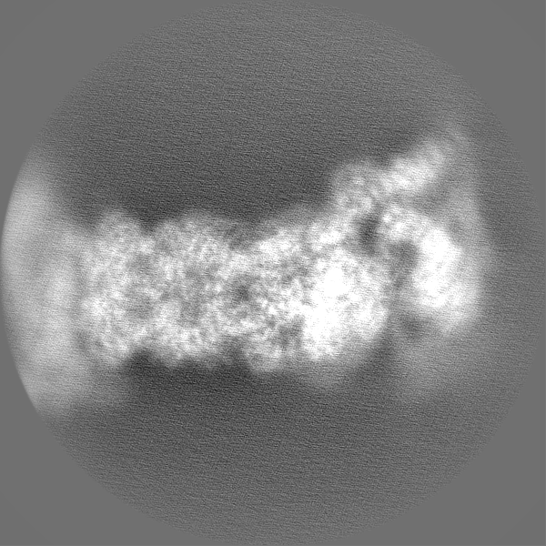
| ムービー |
ムービービューア |
|---|---|
| 構造ビューア | EMマップ: SurfViewMolmilJmol/JSmol |
| 添付画像 |
- ダウンロードとリンク
-EMDBアーカイブ
| マップデータ | emd_9217.map.gz | 748.5 MB | EMDBマップデータ形式 | |
|---|---|---|---|---|
| ヘッダ (付随情報) | emd-9217-v30.xmlemd-9217.xml | 68.9 KB 68.9 KB | 表示 表示 | EMDBヘッダ |
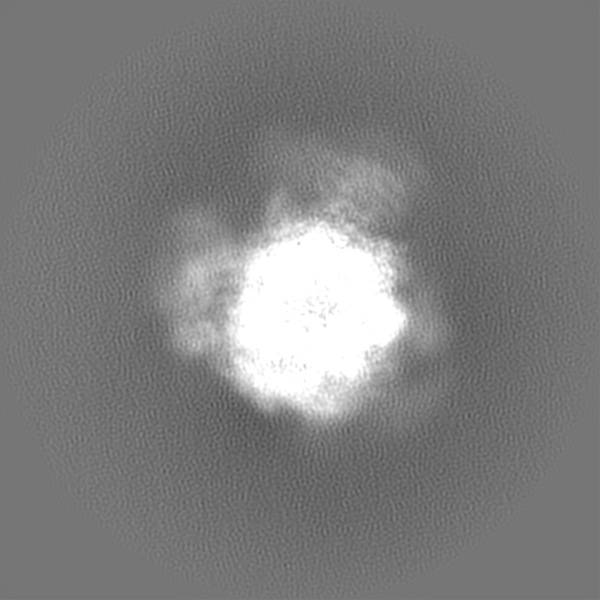
| 画像 |  emd_9217.png emd_9217.png | 37.6 KB | ||
| Filedesc metadata | emd-9217.cif.gz | 15.8 KB | ||
| その他 | emd_9217_additional_1.map.gzemd_9217_additional_2.map.gz | 731.4 MB 756.3 MB | ||
| アーカイブディレクトリ |  http://ftp.pdbj.org/pub/emdb/structures/EMD-9217ftp://ftp.pdbj.org/pub/emdb/structures/EMD-9217 http://ftp.pdbj.org/pub/emdb/structures/EMD-9217ftp://ftp.pdbj.org/pub/emdb/structures/EMD-9217 | HTTPS FTP |
-関連構造データ
| 関連構造データ | 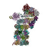 6msdMC 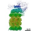 9215C 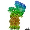 9216C 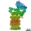 9218C 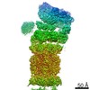 9219C 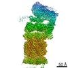 9220C 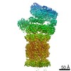 9221C 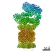 9222C 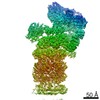 9223C  9224C 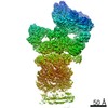 9225C 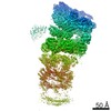 9226C 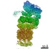 9227C 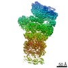 9228C  9229C  6msbC 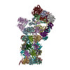 6mseC 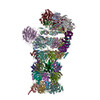 6msgC 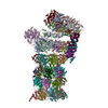 6mshC 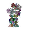 6msjC 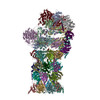 6mskC C: 同じ文献を引用 ( M: このマップから作成された原子モデル |
|---|---|
| 類似構造データ | |
| 電子顕微鏡画像生データ | EMPIAR-10669 (タイトル: Cryo-EM dataset of the substrate-engaged human 26S proteasome Data size: 13.9 TB Data #1: Drift-corrected frame-averaged super-counting mode micrographs and extracted particles of substrate-engaged human 26S proteasome [micrographs - single frame]) |

-リンク
| EMDBのページ | EMDB (EBI/PDBe) / EMDataResource |
|---|---|
| 「今月の分子」の関連する項目 |


-マップ
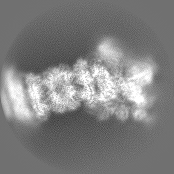
| ファイル | ダウンロード / ファイル: emd_9217.map.gz / 形式: CCP4 / 大きさ: 824 MB / タイプ: IMAGE STORED AS FLOATING POINT NUMBER (4 BYTES) | ||||||||||||||||||||||||||||||||||||||||||||||||||||||||||||||||||||
|---|---|---|---|---|---|---|---|---|---|---|---|---|---|---|---|---|---|---|---|---|---|---|---|---|---|---|---|---|---|---|---|---|---|---|---|---|---|---|---|---|---|---|---|---|---|---|---|---|---|---|---|---|---|---|---|---|---|---|---|---|---|---|---|---|---|---|---|---|---|
| 注釈 | The complete map of state EA2 of substrate-engaged human proteasome, low pass-filtered to 3 Angstrom without amplitude correction. | ||||||||||||||||||||||||||||||||||||||||||||||||||||||||||||||||||||
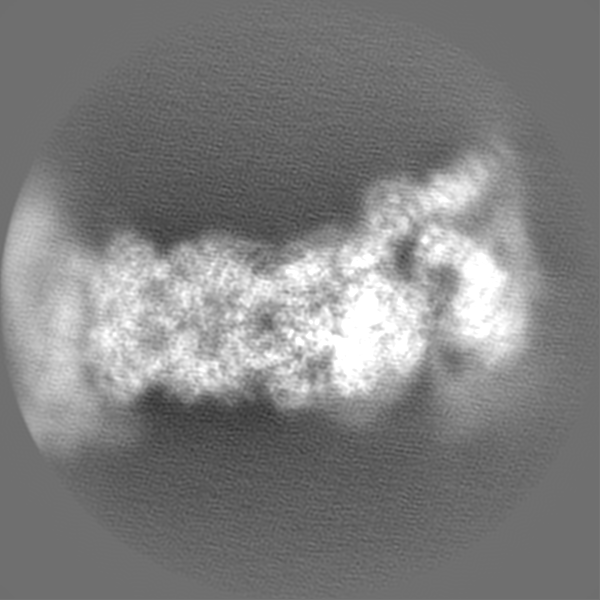
| 投影像・断面図 | 画像のコントロール
画像は Spider により作成 | ||||||||||||||||||||||||||||||||||||||||||||||||||||||||||||||||||||
| ボクセルのサイズ | X=Y=Z: 0.685 Å | ||||||||||||||||||||||||||||||||||||||||||||||||||||||||||||||||||||
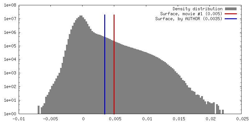
| 密度 |
| ||||||||||||||||||||||||||||||||||||||||||||||||||||||||||||||||||||
| 対称性 | 空間群: 1 | ||||||||||||||||||||||||||||||||||||||||||||||||||||||||||||||||||||
| 詳細 | EMDB XML:
CCP4マップ ヘッダ情報:
| ||||||||||||||||||||||||||||||||||||||||||||||||||||||||||||||||||||
 Z (Sec.)
 Y (Row.)
 X (Col.)


-添付データ
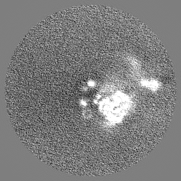
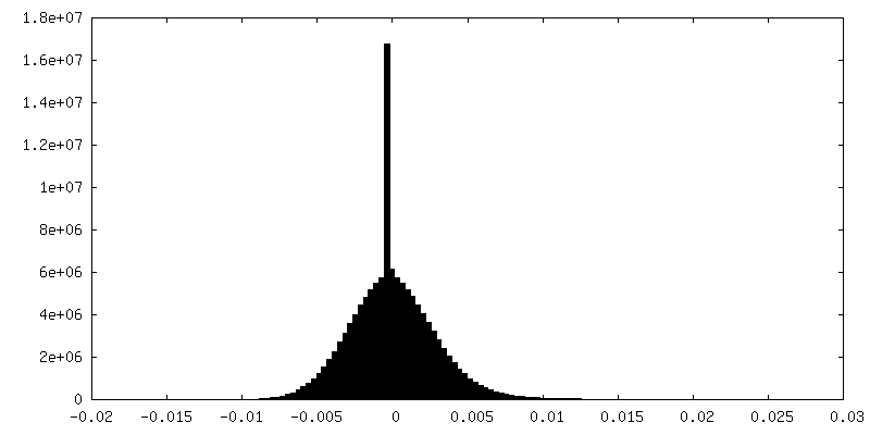
-追加マップ: Unfiltered, uncorrected raw EA2 map of complete holoenzyme
| ファイル | emd_9217_additional_1.map | ||||||||||||
|---|---|---|---|---|---|---|---|---|---|---|---|---|---|
| 注釈 | Unfiltered, uncorrected raw EA2 map of complete holoenzyme | ||||||||||||
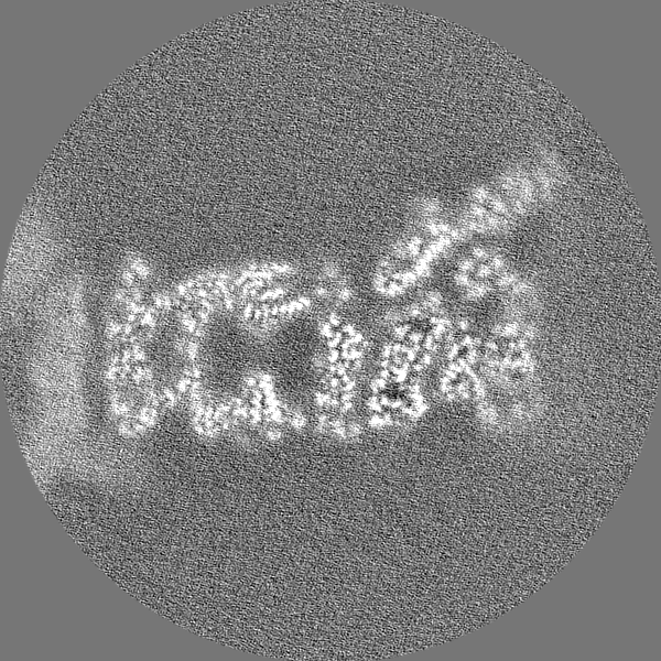
| 投影像・断面図 |
| ||||||||||||
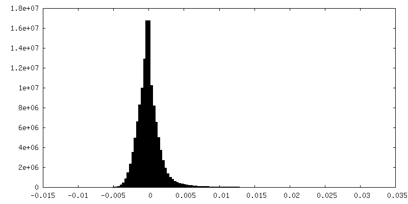
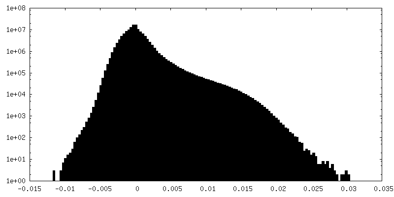
| 密度ヒストグラム |


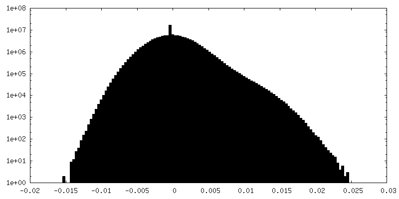
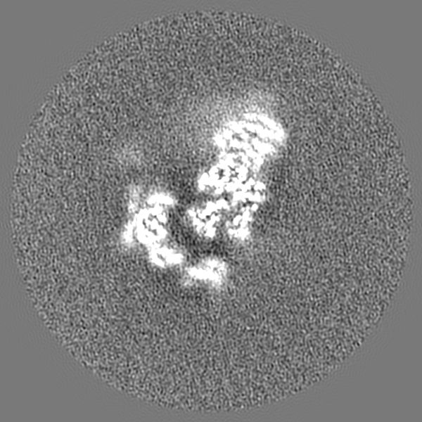
-追加マップ: The complete map of state EA2 of substrate-engaged...
| ファイル | emd_9217_additional_2.map | ||||||||||||
|---|---|---|---|---|---|---|---|---|---|---|---|---|---|
| 注釈 | The complete map of state EA2 of substrate-engaged human proteasome, low pass-filtered to 3.2 Angstrom with amplitude correction with a B-factor of -46 | ||||||||||||
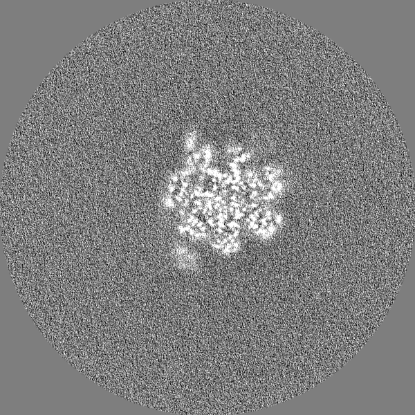
| 投影像・断面図 |
| ||||||||||||
| 密度ヒストグラム |

- 試料の構成要素
+全体 : Human 26S proteasome
+超分子 #1: Human 26S proteasome
+分子 #1: 26S proteasome non-ATPase regulatory subunit 1
+分子 #2: 26S proteasome non-ATPase regulatory subunit 3
+分子 #3: 26S proteasome non-ATPase regulatory subunit 12
+分子 #4: 26S proteasome non-ATPase regulatory subunit 11
+分子 #5: 26S proteasome non-ATPase regulatory subunit 6
+分子 #6: 26S proteasome non-ATPase regulatory subunit 7
+分子 #7: 26S proteasome non-ATPase regulatory subunit 13
+分子 #8: 26S proteasome non-ATPase regulatory subunit 4
+分子 #9: 26S proteasome non-ATPase regulatory subunit 14
+分子 #10: 26S proteasome non-ATPase regulatory subunit 8
+分子 #11: 26S proteasome complex subunit SEM1
+分子 #12: 26S proteasome non-ATPase regulatory subunit 2
+分子 #13: 26S proteasome regulatory subunit 7
+分子 #14: 26S proteasome regulatory subunit 4
+分子 #15: 26S proteasome regulatory subunit 8
+分子 #16: 26S proteasome regulatory subunit 6B
+分子 #17: 26S proteasome regulatory subunit 10B
+分子 #18: 26S proteasome regulatory subunit 6A
+分子 #19: Ubiquitin
+分子 #20: Proteasome subunit alpha type-6
+分子 #21: Proteasome subunit alpha type-2
+分子 #22: Proteasome subunit alpha type-4
+分子 #23: Proteasome subunit alpha type-7
+分子 #24: Proteasome subunit alpha type-5
+分子 #25: Proteasome subunit alpha type-1
+分子 #26: Proteasome subunit alpha type-3
+分子 #27: Proteasome subunit beta type-6
+分子 #28: Proteasome subunit beta type-7
+分子 #29: Proteasome subunit beta type-3
+分子 #30: Proteasome subunit beta type-2
+分子 #31: Proteasome subunit beta type-5
+分子 #32: Proteasome subunit beta type-1
+分子 #33: Proteasome subunit beta type-4
+分子 #34: ZINC ION
+分子 #35: ADENOSINE-5'-TRIPHOSPHATE

+分子 #36: MAGNESIUM ION
+分子 #37: ADENOSINE-5'-DIPHOSPHATE

-実験情報
-構造解析
| 手法 | クライオ電子顕微鏡法 |
|---|---|
 解析 解析 | 単粒子再構成法 |
| 試料の集合状態 | particle |
-試料調製
| 緩衝液 | pH: 8 |
|---|---|
| グリッド | 詳細: unspecified |
| 凍結 | 凍結剤: ETHANE |
- 電子顕微鏡法
| 顕微鏡 | FEI TITAN KRIOS |
|---|---|
| 撮影 | フィルム・検出器のモデル: GATAN K2 SUMMIT (4k x 4k) 平均電子線量: 44.0 e/Å2 |
| 電子線 | 加速電圧: 300 kV / 電子線源:  FIELD EMISSION GUN FIELD EMISSION GUN |
| 電子光学系 | 照射モード: FLOOD BEAM / 撮影モード: BRIGHT FIELD |
| 実験機器 |  モデル: Titan Krios / 画像提供: FEI Company |
