 ムービー
ムービー コントローラー
 構造ビューア
 万見文献について


+検索条件
-Structure paper
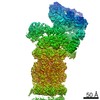
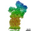
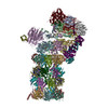
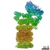
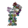
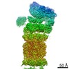
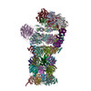
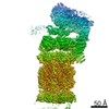
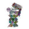
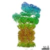
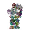
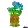
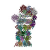
| タイトル | Cryo-EM structures and dynamics of substrate-engaged human 26S proteasome. |
|---|---|
| ジャーナル・号・ページ | Nature, Vol. 565, Issue 7737, Page 49-55, Year 2019 |
| 掲載日 | 2018年11月12日 |
 著者 著者 | Yuanchen Dong / Shuwen Zhang / Zhaolong Wu / Xuemei Li / Wei Li Wang / Yanan Zhu / Svetla Stoilova-McPhie / Ying Lu / Daniel Finley / Youdong Mao /   |
| PubMed 要旨 | The proteasome is an ATP-dependent, 2.5-megadalton molecular machine that is responsible for selective protein degradation in eukaryotic cells. Here we present cryo-electron microscopy structures of ...The proteasome is an ATP-dependent, 2.5-megadalton molecular machine that is responsible for selective protein degradation in eukaryotic cells. Here we present cryo-electron microscopy structures of the substrate-engaged human proteasome in seven conformational states at 2.8-3.6 Å resolution, captured during breakdown of a polyubiquitylated protein. These structures illuminate a spatiotemporal continuum of dynamic substrate-proteasome interactions from ubiquitin recognition to substrate translocation, during which ATP hydrolysis sequentially navigates through all six ATPases. There are three principal modes of coordinated hydrolysis, featuring hydrolytic events in two oppositely positioned ATPases, in two adjacent ATPases and in one ATPase at a time. These hydrolytic modes regulate deubiquitylation, initiation of translocation and processive unfolding of substrates, respectively. Hydrolysis of ATP powers a hinge-like motion in each ATPase that regulates its substrate interaction. Synchronization of ATP binding, ADP release and ATP hydrolysis in three adjacent ATPases drives rigid-body rotations of substrate-bound ATPases that are propagated unidirectionally in the ATPase ring and unfold the substrate. |
 リンク リンク | Nature / PubMed:30479383 / PubMed Central |
| 手法 | EM (単粒子) |
| 解像度 | 2.8 - 3.6 Å |
| 構造データ | 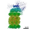 EMDB-9215: EMDB-9216, PDB-6msb: EMDB-9217, PDB-6msd: EMDB-9218, PDB-6mse: EMDB-9219, PDB-6msg: EMDB-9220, PDB-6msh: EMDB-9221, PDB-6msj: EMDB-9222, PDB-6msk: 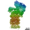 EMDB-9223:  EMDB-9224: 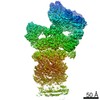 EMDB-9225: 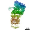 EMDB-9226: 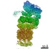 EMDB-9227: 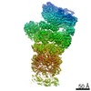 EMDB-9228:  EMDB-9229: |
| 化合物 |  ChemComp-ZN:  ChemComp-ATP:  ChemComp-MG:  ChemComp-ADP: |
| 由来 |
|
 キーワード キーワード | HYDROLASE / Proteosome |

 homo sapiens (ヒト)
