 Movie
Movie Controller

[English] 日本語
 Yorodumi
- PDB-6msd: Cryo-EM structures and dynamics of substrate-engaged human 26S pr... -
+ Open data

- Basic information
| Entry | Database: PDB / ID: 6msd | |||||||||||||||||||||||||||||||||||||||||||||||||||||||||||||||||||||
|---|---|---|---|---|---|---|---|---|---|---|---|---|---|---|---|---|---|---|---|---|---|---|---|---|---|---|---|---|---|---|---|---|---|---|---|---|---|---|---|---|---|---|---|---|---|---|---|---|---|---|---|---|---|---|---|---|---|---|---|---|---|---|---|---|---|---|---|---|---|---|
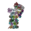
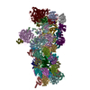
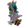
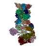
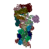
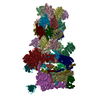
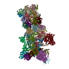
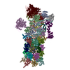
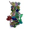
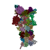
| Title | Cryo-EM structures and dynamics of substrate-engaged human 26S proteasome | |||||||||||||||||||||||||||||||||||||||||||||||||||||||||||||||||||||
 Components Components |
| |||||||||||||||||||||||||||||||||||||||||||||||||||||||||||||||||||||
 Keywords Keywords | HYDROLASE / Proteosome | |||||||||||||||||||||||||||||||||||||||||||||||||||||||||||||||||||||
| Function / homology |  Function and homology information Function and homology informationthyrotropin-releasing hormone receptor binding / nuclear proteasome complex / host-mediated perturbation of viral transcription / positive regulation of inclusion body assembly / Impaired BRCA2 translocation to the nucleus / Impaired BRCA2 binding to SEM1 (DSS1) / proteasome accessory complex / purine ribonucleoside triphosphate binding / integrator complex / proteasome regulatory particle ...thyrotropin-releasing hormone receptor binding / nuclear proteasome complex / host-mediated perturbation of viral transcription / positive regulation of inclusion body assembly / Impaired BRCA2 translocation to the nucleus / Impaired BRCA2 binding to SEM1 (DSS1) / proteasome accessory complex / purine ribonucleoside triphosphate binding / integrator complex / proteasome regulatory particle / CD8-positive, alpha-beta T cell differentiation / thymic T cell selection / CD8-positive, alpha-beta T cell homeostasis / cytosolic proteasome complex / positive regulation of proteasomal protein catabolic process / hypothalamus gonadotrophin-releasing hormone neuron development / proteasome-activating activity / Antigen processing: Ub, ATP-independent proteasomal degradation / female meiosis I / proteasome regulatory particle, lid subcomplex / proteasome regulatory particle, base subcomplex / seminiferous tubule development / positive regulation of protein monoubiquitination / fat pad development / negative regulation of regulatory T cell differentiation / mitochondrion transport along microtubule / T-helper 1 cell differentiation / protein K63-linked deubiquitination / metal-dependent deubiquitinase activity / cellular response to type I interferon / negative regulation of programmed cell death / Regulation of ornithine decarboxylase (ODC) / proteasome core complex / Proteasome assembly / T-helper 17 cell differentiation / retrograde vesicle-mediated transport, Golgi to endoplasmic reticulum / Cross-presentation of soluble exogenous antigens (endosomes) / transcription factor binding / K63-linked deubiquitinase activity / Somitogenesis / flagellated sperm motility / Homologous DNA Pairing and Strand Exchange / Defective homologous recombination repair (HRR) due to BRCA1 loss of function / Defective HDR through Homologous Recombination Repair (HRR) due to PALB2 loss of BRCA1 binding function / Defective HDR through Homologous Recombination Repair (HRR) due to PALB2 loss of BRCA2/RAD51/RAD51C binding function / Resolution of D-loop Structures through Synthesis-Dependent Strand Annealing (SDSA) / female gonad development / Resolution of D-loop Structures through Holliday Junction Intermediates / proteasome binding / Impaired BRCA2 binding to RAD51 / myofibril / male meiosis I / positive regulation of RNA polymerase II transcription preinitiation complex assembly / proteasomal ubiquitin-independent protein catabolic process / general transcription initiation factor binding / Presynaptic phase of homologous DNA pairing and strand exchange / proteasome storage granule / positive regulation of intrinsic apoptotic signaling pathway by p53 class mediator / AMPK-induced ERAD and lysosome mediated degradation of PD-L1(CD274) / protein deubiquitination / polyubiquitin modification-dependent protein binding / proteasome endopeptidase complex / NF-kappaB binding / proteasome core complex, beta-subunit complex / endopeptidase activator activity / energy homeostasis / threonine-type endopeptidase activity / GSK3B-mediated proteasomal degradation of PD-L1(CD274) / proteasome core complex, alpha-subunit complex / SPOP-mediated proteasomal degradation of PD-L1(CD274) / proteasome assembly / mRNA export from nucleus / SARS-CoV-1 targets host intracellular signalling and regulatory pathways / immune system process / regulation of G1/S transition of mitotic cell cycle / regulation of macroautophagy / enzyme regulator activity / Ribosome Quality Control (RQC) complex extracts and degrades nascent peptide / neuron projection morphogenesis / positive regulation of interleukin-2 production / ciliary tip / ERAD pathway / response to type II interferon / inclusion body / Maturation of protein E / Maturation of protein E / ER Quality Control Compartment (ERQC) / regulation of neuron apoptotic process / Myoclonic epilepsy of Lafora / FLT3 signaling by CBL mutants / IRAK2 mediated activation of TAK1 complex / Alpha-protein kinase 1 signaling pathway / Glycogen synthesis / IRAK1 recruits IKK complex / IRAK1 recruits IKK complex upon TLR7/8 or 9 stimulation / Prevention of phagosomal-lysosomal fusion / Endosomal Sorting Complex Required For Transport (ESCRT) / Membrane binding and targetting of GAG proteins / Regulation of TBK1, IKKε (IKBKE)-mediated activation of IRF3, IRF7 / Negative regulation of FLT3 Similarity search - Function | |||||||||||||||||||||||||||||||||||||||||||||||||||||||||||||||||||||
| Biological species |  Homo sapiens (human) Homo sapiens (human) | |||||||||||||||||||||||||||||||||||||||||||||||||||||||||||||||||||||
| Method | ELECTRON MICROSCOPY / single particle reconstruction / cryo EM / Resolution: 3.2 Å | |||||||||||||||||||||||||||||||||||||||||||||||||||||||||||||||||||||
 Authors Authors | Mao, Y.D. | |||||||||||||||||||||||||||||||||||||||||||||||||||||||||||||||||||||
| Funding support |  China, China,  United States, 3items United States, 3items
| |||||||||||||||||||||||||||||||||||||||||||||||||||||||||||||||||||||
 Citation Citation | Journal: Nature / Year: 2019 Title: Cryo-EM structures and dynamics of substrate-engaged human 26S proteasome. Authors: Yuanchen Dong / Shuwen Zhang / Zhaolong Wu / Xuemei Li / Wei Li Wang / Yanan Zhu / Svetla Stoilova-McPhie / Ying Lu / Daniel Finley / Youdong Mao / Abstract: The proteasome is an ATP-dependent, 2.5-megadalton molecular machine that is responsible for selective protein degradation in eukaryotic cells. Here we present cryo-electron microscopy structures of ...The proteasome is an ATP-dependent, 2.5-megadalton molecular machine that is responsible for selective protein degradation in eukaryotic cells. Here we present cryo-electron microscopy structures of the substrate-engaged human proteasome in seven conformational states at 2.8-3.6 Å resolution, captured during breakdown of a polyubiquitylated protein. These structures illuminate a spatiotemporal continuum of dynamic substrate-proteasome interactions from ubiquitin recognition to substrate translocation, during which ATP hydrolysis sequentially navigates through all six ATPases. There are three principal modes of coordinated hydrolysis, featuring hydrolytic events in two oppositely positioned ATPases, in two adjacent ATPases and in one ATPase at a time. These hydrolytic modes regulate deubiquitylation, initiation of translocation and processive unfolding of substrates, respectively. Hydrolysis of ATP powers a hinge-like motion in each ATPase that regulates its substrate interaction. Synchronization of ATP binding, ADP release and ATP hydrolysis in three adjacent ATPases drives rigid-body rotations of substrate-bound ATPases that are propagated unidirectionally in the ATPase ring and unfold the substrate. | |||||||||||||||||||||||||||||||||||||||||||||||||||||||||||||||||||||
| History |
|
- Structure visualization
| Movie |
Movie viewer |
|---|---|
| Structure viewer | Molecule: MolmilJmol/JSmol |
- Downloads & links
-Download
| PDBx/mmCIF format | 6msd.cif.gz | 2.3 MB | Display | PDBx/mmCIF format |
|---|---|---|---|---|
| PDB format | pdb6msd.ent.gz | Display | PDB format | |
| PDBx/mmJSON format | 6msd.json.gz | Tree view | PDBx/mmJSON format | |
| Others |  Other downloads Other downloads |
-Validation report
| Arichive directory | https://data.pdbj.org/pub/pdb/validation_reports/ms/6msdftp://data.pdbj.org/pub/pdb/validation_reports/ms/6msd | HTTPS FTP |
|---|
-Related structure data
| Related structure data | 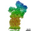 9217MC 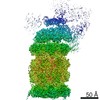 9215C 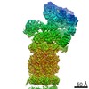 9216C 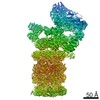 9218C 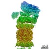 9219C 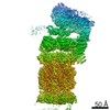 9220C 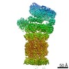 9221C 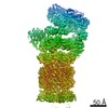 9222C 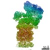 9223C  9224C 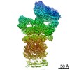 9225C 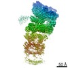 9226C 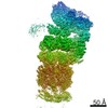 9227C 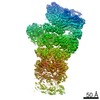 9228C  9229C  6msbC 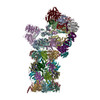 6mseC 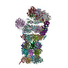 6msgC 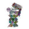 6mshC 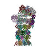 6msjC 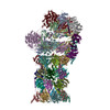 6mskC M: map data used to model this data C: citing same article ( |
|---|---|
| Similar structure data | |
| EM raw data | EMPIAR-10669 (Title: Cryo-EM dataset of the substrate-engaged human 26S proteasome Data size: 13.9 TB Data #1: Drift-corrected frame-averaged super-counting mode micrographs and extracted particles of substrate-engaged human 26S proteasome [micrographs - single frame]) |

-Links
 PDBj
PDBj


- Assembly
| Deposited unit | 
|
|---|---|
| 1 |
|
-Components
-26S proteasome non-ATPase regulatory subunit ... , 11 types, 11 molecules UVWXYZabcdf
| #1: Protein | Mass: 105958.234 Da / Num. of mol.: 1 Source method: isolated from a genetically manipulated source Source: (gene. exp.) Homo sapiens (human) / Gene: PSMD1 / Production host: Homo sapiens (human) / References: UniProt: Q99460 |
|---|---|
| #2: Protein | Mass: 61066.500 Da / Num. of mol.: 1 Source method: isolated from a genetically manipulated source Source: (gene. exp.) Homo sapiens (human) / Gene: PSMD3 / Production host: Homo sapiens (human) / References: UniProt: O43242 |
| #3: Protein | Mass: 52979.359 Da / Num. of mol.: 1 Source method: isolated from a genetically manipulated source Source: (gene. exp.) Homo sapiens (human) / Gene: PSMD12 / Production host: Homo sapiens (human) / References: UniProt: O00232 |
| #4: Protein | Mass: 47526.688 Da / Num. of mol.: 1 Source method: isolated from a genetically manipulated source Source: (gene. exp.) Homo sapiens (human) / Gene: PSMD11 / Production host: Homo sapiens (human) / References: UniProt: O00231 |
| #5: Protein | Mass: 45592.285 Da / Num. of mol.: 1 Source method: isolated from a genetically manipulated source Source: (gene. exp.) Homo sapiens (human) / Gene: PSMD6, KIAA0107, PFAAP4 / Production host: Homo sapiens (human) / References: UniProt: Q15008 |
| #6: Protein | Mass: 37086.441 Da / Num. of mol.: 1 Source method: isolated from a genetically manipulated source Source: (gene. exp.) Homo sapiens (human) / Gene: PSMD7, MOV34L / Production host: Homo sapiens (human) / References: UniProt: P51665 |
| #7: Protein | Mass: 42995.359 Da / Num. of mol.: 1 Source method: isolated from a genetically manipulated source Source: (gene. exp.) Homo sapiens (human) / Gene: PSMD13 / Production host: Homo sapiens (human) / References: UniProt: Q9UNM6 |
| #8: Protein | Mass: 40781.590 Da / Num. of mol.: 1 Source method: isolated from a genetically manipulated source Source: (gene. exp.) Homo sapiens (human) / Gene: PSMD4, MCB1 / Production host: Homo sapiens (human) / References: UniProt: P55036 |
| #9: Protein | Mass: 34488.824 Da / Num. of mol.: 1 Source method: isolated from a genetically manipulated source Source: (gene. exp.) Homo sapiens (human) / Gene: PSMD14, POH1 / Production host: Homo sapiens (human)References: UniProt: O00487, Hydrolases; Acting on peptide bonds (peptidases); Omega peptidases |
| #10: Protein | Mass: 39536.676 Da / Num. of mol.: 1 Source method: isolated from a genetically manipulated source Source: (gene. exp.) Homo sapiens (human) / Gene: PSMD8 / Production host: Homo sapiens (human) / References: UniProt: P48556 |
| #12: Protein | Mass: 100313.625 Da / Num. of mol.: 1 Source method: isolated from a genetically manipulated source Source: (gene. exp.) Homo sapiens (human) / Gene: PSMD2, TRAP2 / Production host: Homo sapiens (human) / References: UniProt: Q13200 |
-Protein , 2 types, 3 molecules euw
| #11: Protein | Mass: 8284.611 Da / Num. of mol.: 1 Source method: isolated from a genetically manipulated source Source: (gene. exp.) Homo sapiens (human) / Gene: SEM1, C7orf76, DSS1, SHFDG1, SHFM1 / Production host: Homo sapiens (human) / References: UniProt: P60896 |
|---|---|
| #19: Protein | Mass: 8604.845 Da / Num. of mol.: 2 Source method: isolated from a genetically manipulated source Source: (gene. exp.) Homo sapiens (human) / Production host: Homo sapiens (human) / References: UniProt: P0CG47 |
-26S proteasome regulatory subunit ... , 6 types, 6 molecules ABCDEF
| #13: Protein | Mass: 48700.805 Da / Num. of mol.: 1 Source method: isolated from a genetically manipulated source Source: (gene. exp.) Homo sapiens (human) / Gene: PSMC2, MSS1 / Production host: Homo sapiens (human) / References: UniProt: P35998 |
|---|---|
| #14: Protein | Mass: 49260.504 Da / Num. of mol.: 1 Source method: isolated from a genetically manipulated source Source: (gene. exp.) Homo sapiens (human) / Gene: PSMC1 / Production host: Homo sapiens (human) / References: UniProt: P62191 |
| #15: Protein | Mass: 44852.121 Da / Num. of mol.: 1 Source method: isolated from a genetically manipulated source Source: (gene. exp.) Homo sapiens (human) / Gene: PSMC5, SUG1 / Production host: Homo sapiens (human) / References: UniProt: P62195 |
| #16: Protein | Mass: 47426.141 Da / Num. of mol.: 1 Source method: isolated from a genetically manipulated source Source: (gene. exp.) Homo sapiens (human) / Gene: PSMC4, MIP224, TBP7 / Production host: Homo sapiens (human) / References: UniProt: P43686 |
| #17: Protein | Mass: 45867.027 Da / Num. of mol.: 1 Source method: isolated from a genetically manipulated source Source: (gene. exp.) Homo sapiens (human) / Gene: PSMC6 / Production host: Homo sapiens (human) / References: UniProt: A0A087X2I1, UniProt: P62333*PLUS |
| #18: Protein | Mass: 49266.457 Da / Num. of mol.: 1 Source method: isolated from a genetically manipulated source Source: (gene. exp.) Homo sapiens (human) / Gene: PSMC3, TBP1 / Production host: Homo sapiens (human) / References: UniProt: P17980 |
-Proteasome subunit alpha type- ... , 7 types, 14 molecules GgHhIiJjKkLlMm
| #20: Protein | Mass: 27301.262 Da / Num. of mol.: 2 Source method: isolated from a genetically manipulated source Source: (gene. exp.) Homo sapiens (human) / Gene: PSMA6, PROS27 / Production host: Homo sapiens (human)References: UniProt: P60900, proteasome endopeptidase complex #21: Protein | Mass: 25796.338 Da / Num. of mol.: 2 Source method: isolated from a genetically manipulated source Source: (gene. exp.) Homo sapiens (human) / Gene: PSMA2, HC3, PSC3 / Production host: Homo sapiens (human)References: UniProt: P25787, proteasome endopeptidase complex #22: Protein | Mass: 29394.648 Da / Num. of mol.: 2 Source method: isolated from a genetically manipulated source Source: (gene. exp.) Homo sapiens (human) / Gene: PSMA4, HC9, PSC9 / Production host: Homo sapiens (human)References: UniProt: P25789, proteasome endopeptidase complex #23: Protein | Mass: 27798.695 Da / Num. of mol.: 2 Source method: isolated from a genetically manipulated source Source: (gene. exp.) Homo sapiens (human) / Gene: PSMA7, HSPC / Production host: Homo sapiens (human)References: UniProt: O14818, proteasome endopeptidase complex #24: Protein | Mass: 26304.779 Da / Num. of mol.: 2 Source method: isolated from a genetically manipulated source Source: (gene. exp.) Homo sapiens (human) / Gene: PSMA5 / Production host: Homo sapiens (human)References: UniProt: P28066, proteasome endopeptidase complex #25: Protein | Mass: 30150.277 Da / Num. of mol.: 2 Source method: isolated from a genetically manipulated source Source: (gene. exp.) Homo sapiens (human) / Gene: PSMA1, HC2, NU, PROS30, PSC2 / Production host: Homo sapiens (human)References: UniProt: P25786, proteasome endopeptidase complex #26: Protein | Mass: 28338.057 Da / Num. of mol.: 2 Source method: isolated from a genetically manipulated source Source: (gene. exp.) Homo sapiens (human) / Gene: PSMA3, HC8, PSC8 / Production host: Homo sapiens (human)References: UniProt: P25788, proteasome endopeptidase complex |
|---|
-Proteasome subunit beta type- ... , 7 types, 14 molecules NnOoPpQqRrSsTt
| #27: Protein | Mass: 25246.455 Da / Num. of mol.: 2 Source method: isolated from a genetically manipulated source Source: (gene. exp.) Homo sapiens (human) / Gene: PSMB6, LMPY, Y / Production host: Homo sapiens (human)References: UniProt: P28072, proteasome endopeptidase complex #28: Protein | Mass: 29869.223 Da / Num. of mol.: 2 Source method: isolated from a genetically manipulated source Source: (gene. exp.) Homo sapiens (human) / Gene: PSMB7, Z / Production host: Homo sapiens (human)References: UniProt: Q99436, proteasome endopeptidase complex #29: Protein | Mass: 22841.701 Da / Num. of mol.: 2 Source method: isolated from a genetically manipulated source Source: (gene. exp.) Homo sapiens (human) / Gene: PSMB3 / Production host: Homo sapiens (human)References: UniProt: P49720, proteasome endopeptidase complex #30: Protein | Mass: 22864.277 Da / Num. of mol.: 2 Source method: isolated from a genetically manipulated source Source: (gene. exp.) Homo sapiens (human) / Gene: PSMB2 / Production host: Homo sapiens (human)References: UniProt: P49721, proteasome endopeptidase complex #31: Protein | Mass: 28379.053 Da / Num. of mol.: 2 Source method: isolated from a genetically manipulated source Source: (gene. exp.) Homo sapiens (human) / Gene: PSMB5, LMPX, MB1, X / Production host: Homo sapiens (human)References: UniProt: P28074, proteasome endopeptidase complex #32: Protein | Mass: 26391.201 Da / Num. of mol.: 2 Source method: isolated from a genetically manipulated source Source: (gene. exp.) Homo sapiens (human) / Gene: PSMB1, PSC5 / Production host: Homo sapiens (human)References: UniProt: P20618, proteasome endopeptidase complex #33: Protein | Mass: 29099.986 Da / Num. of mol.: 2 Source method: isolated from a genetically manipulated source Source: (gene. exp.) Homo sapiens (human) / Gene: PSMB4, PROS26 / Production host: Homo sapiens (human)References: UniProt: P28070, proteasome endopeptidase complex |
|---|
-Non-polymers , 4 types, 12 molecules 


| #34: Chemical | ChemComp-ZN /  Mass: 65.409 Da / Num. of mol.: 1 / Source method: obtained synthetically / Formula: Zn Mass: 65.409 Da / Num. of mol.: 1 / Source method: obtained synthetically / Formula: Zn | ||||
|---|---|---|---|---|---|
| #35: Chemical | ChemComp-ATP /  Mass: 507.181 Da / Num. of mol.: 4 / Source method: obtained synthetically / Formula: C10H16N5O13P3 / Comment: ATP, energy-carrying molecule*YM Mass: 507.181 Da / Num. of mol.: 4 / Source method: obtained synthetically / Formula: C10H16N5O13P3 / Comment: ATP, energy-carrying molecule*YM#36: Chemical | ChemComp-MG /  Mass: 24.305 Da / Num. of mol.: 5 / Source method: obtained synthetically / Formula: Mg Mass: 24.305 Da / Num. of mol.: 5 / Source method: obtained synthetically / Formula: Mg#37: Chemical |  Mass: 427.201 Da / Num. of mol.: 2 / Source method: obtained synthetically / Formula: C10H15N5O10P2 / Comment: ADP, energy-carrying molecule*YM Mass: 427.201 Da / Num. of mol.: 2 / Source method: obtained synthetically / Formula: C10H15N5O10P2 / Comment: ADP, energy-carrying molecule*YM |
-Details
| Has protein modification | Y |
|---|
-Experimental details
-Experiment
| Experiment | Method: ELECTRON MICROSCOPY |
|---|---|
| EM experiment | Aggregation state: PARTICLE / 3D reconstruction method: single particle reconstruction |
- Sample preparation
| Component | Name: Human 26S proteasome / Type: COMPLEX / Entity ID: #1-#33 / Source: RECOMBINANT |
|---|---|
| Source (natural) | Organism: Homo sapiens (human) |
| Source (recombinant) | Organism: Homo sapiens (human) |
| Buffer solution | pH: 8 |
| Specimen | Embedding applied: NO / Shadowing applied: NO / Staining applied: NO / Vitrification applied: YES |
| Specimen support | Details: unspecified |
| Vitrification | Cryogen name: ETHANE |
- Electron microscopy imaging
| Experimental equipment |  Model: Titan Krios / Image courtesy: FEI Company |
|---|---|
| Microscopy | Model: FEI TITAN KRIOS |
| Electron gun | Electron source:  FIELD EMISSION GUN / Accelerating voltage: 300 kV / Illumination mode: FLOOD BEAM FIELD EMISSION GUN / Accelerating voltage: 300 kV / Illumination mode: FLOOD BEAM |
| Electron lens | Mode: BRIGHT FIELD |
| Image recording | Electron dose: 44 e/Å2 / Film or detector model: GATAN K2 SUMMIT (4k x 4k) |
- Processing
| Software | Name: PHENIX / Version: 1.13_2998: / Classification: refinement |
|---|---|
| EM software | Name: PHENIX / Category: model refinement |
| CTF correction | Type: PHASE FLIPPING AND AMPLITUDE CORRECTION |
| Symmetry | Point symmetry: C1 (asymmetric) |
| 3D reconstruction | Resolution: 3.2 Å / Resolution method: FSC 0.143 CUT-OFF / Num. of particles: 79691 / Symmetry type: POINT |
