 Movie
Movie Controller

[English] 日本語
 Yorodumi
- EMDB-9040: Analysis of bacteriophage phiX174 tomograms using the locked self... -
+ Open data

- Basic information
| Entry | Database: EMDB / ID: EMD-9040 | ||||||||||||
|---|---|---|---|---|---|---|---|---|---|---|---|---|---|
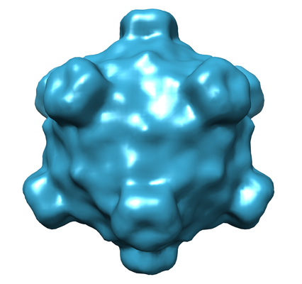
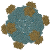
| Title | Analysis of bacteriophage phiX174 tomograms using the locked self-rotation function | ||||||||||||
 Map data Map data | The map was calculated by averaging of ten icosahedrally-averaged maps of individual phiX174 particles. | ||||||||||||
 Sample Sample |
| ||||||||||||
| Biological species |  Enterobacteria phage phiX174 (virus) Enterobacteria phage phiX174 (virus) | ||||||||||||
| Method | electron tomography / cryo EM | ||||||||||||
 Authors Authors | Fokine A / Sun Y / Khare B / Rossmann MG | ||||||||||||
| Funding support |  United States, 3 items United States, 3 items
| ||||||||||||
 Citation Citation | Journal: J Struct Biol / Year: 2019 Title: Enhancement of tomogram interpretability using the locked self-rotation function. Authors: Andrei Fokine / Baldeep Khare / Yingyuan Sun / Michael G Rossmann / Abstract: The interpretation of cryo-electron tomograms of macromolecular complexes can be difficult because of the large amount of noise and because of the missing wedge effect. Here it is shown how the ...The interpretation of cryo-electron tomograms of macromolecular complexes can be difficult because of the large amount of noise and because of the missing wedge effect. Here it is shown how the presence of rotational symmetry in a sample can be utilized to enhance the quality of a tomographic analysis. The orientation of symmetry axes in a sub-tomogram can be determined using a locked self-rotation function. Given this knowledge, the sub-tomogram density can then be averaged to improve its interpretability. Sub-tomograms of the icosahedral bacteriophage phiX174 are used to demonstrate the procedure. | ||||||||||||
| History |
|
- Structure visualization
| Movie |
 Movie viewer Movie viewer |
|---|---|
| Structure viewer | EM map: SurfViewMolmilJmol/JSmol |
| Supplemental images |
- Downloads & links
-EMDB archive
| Map data | emd_9040.map.gz | 474.2 KB | EMDB map data format | |
|---|---|---|---|---|
| Header (meta data) | emd-9040-v30.xmlemd-9040.xml | 33.7 KB 33.7 KB | Display Display | EMDB header |
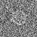
| Images |  emd_9040.png emd_9040.png | 106.8 KB | ||
| Others | emd_9040_additional_1.map.gzemd_9040_additional_10.map.gzemd_9040_additional_11.map.gzemd_9040_additional_2.map.gzemd_9040_additional_3.map.gzemd_9040_additional_4.map.gzemd_9040_additional_5.map.gzemd_9040_additional_6.map.gzemd_9040_additional_7.map.gzemd_9040_additional_8.map.gzemd_9040_additional_9.map.gz | 1.8 MB 1.8 MB 518.5 KB 1.8 MB 1.8 MB 1.8 MB 1.8 MB 7.4 MB 1.8 MB 1.8 MB 1.8 MB | ||
| Archive directory |  http://ftp.pdbj.org/pub/emdb/structures/EMD-9040ftp://ftp.pdbj.org/pub/emdb/structures/EMD-9040 http://ftp.pdbj.org/pub/emdb/structures/EMD-9040ftp://ftp.pdbj.org/pub/emdb/structures/EMD-9040 | HTTPS FTP |
-Validation report
| Summary document | emd_9040_validation.pdf.gz | 78.2 KB | Display | EMDB validaton report |
|---|---|---|---|---|
| Full document | emd_9040_full_validation.pdf.gz | 77.3 KB | Display | |
| Data in XML | emd_9040_validation.xml.gz | 497 B | Display | |
| Arichive directory | https://ftp.pdbj.org/pub/emdb/validation_reports/EMD-9040ftp://ftp.pdbj.org/pub/emdb/validation_reports/EMD-9040 | HTTPS FTP |
-Related structure data
| Similar structure data |
|---|
-Links
| EMDB pages | EMDB (EBI/PDBe) / EMDataResource |
|---|
-Map
| File | Download / File: emd_9040.map.gz / Format: CCP4 / Size: 2 MB / Type: IMAGE STORED AS FLOATING POINT NUMBER (4 BYTES) | ||||||||||||||||||||||||||||||||||||||||||||||||||||||||||||||||||||
|---|---|---|---|---|---|---|---|---|---|---|---|---|---|---|---|---|---|---|---|---|---|---|---|---|---|---|---|---|---|---|---|---|---|---|---|---|---|---|---|---|---|---|---|---|---|---|---|---|---|---|---|---|---|---|---|---|---|---|---|---|---|---|---|---|---|---|---|---|---|
| Annotation | The map was calculated by averaging of ten icosahedrally-averaged maps of individual phiX174 particles. | ||||||||||||||||||||||||||||||||||||||||||||||||||||||||||||||||||||
| Voxel size | X=Y=Z: 5.2 Å | ||||||||||||||||||||||||||||||||||||||||||||||||||||||||||||||||||||
| Density |
| ||||||||||||||||||||||||||||||||||||||||||||||||||||||||||||||||||||
| Symmetry | Space group: 1 | ||||||||||||||||||||||||||||||||||||||||||||||||||||||||||||||||||||
| Details | EMDB XML:
CCP4 map header:
| ||||||||||||||||||||||||||||||||||||||||||||||||||||||||||||||||||||
-Supplemental data
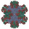
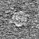
+Additional map: Tomogram of one bacteriophage phiX174 particle.
 Z
Z Y
Y X
X


+Additional map: Tomogram of one bacteriophage phiX174 particle.


+Additional map: The map was produced by averaging of eight...


+Additional map: Tomogram of one bacteriophage phiX174 particle.


+Additional map: Tomogram of one bacteriophage phiX174 particle.


+Additional map: Tomogram of one bacteriophage phiX174 particle.


+Additional map: Tomogram of one bacteriophage phiX174 particle.


+Additional map: Tomogram of one bacteriophage phiX174 particle.
+Additional map: Tomogram of one bacteriophage phiX174 particle.


+Additional map: Tomogram of one bacteriophage phiX174 particle.


+Additional map: Tomogram of one bacteriophage phiX174 particle.


- Sample components
-Entire : Enterobacteria phage phiX174
| Entire | Name: Enterobacteria phage phiX174 (virus) |
|---|---|
| Components |
|
-Supramolecule #1: Enterobacteria phage phiX174
| Supramolecule | Name: Enterobacteria phage phiX174 / type: virus / ID: 1 / Parent: 0 / Details: The virus was grown in E.coli cells. / NCBI-ID: 10847 / Sci species name: Enterobacteria phage phiX174 / Virus type: VIRION / Virus isolate: STRAIN / Virus enveloped: No / Virus empty: No |
|---|---|
| Host (natural) | Organism:  |
| Virus shell | Shell ID: 1 / Diameter: 320.0 Å / T number (triangulation number): 1 |
-Experimental details
-Structure determination
| Method | cryo EM |
|---|---|
 Processing Processing | electron tomography |
| Aggregation state | particle |
-Sample preparation
| Buffer | pH: 7.4 Component:
| ||||||||||||||
|---|---|---|---|---|---|---|---|---|---|---|---|---|---|---|---|
| Grid | Model: C-flat-2/2 4C / Material: COPPER / Pretreatment - Type: GLOW DISCHARGE | ||||||||||||||
| Vitrification | Cryogen name: ETHANE / Instrument: GATAN CRYOPLUNGE 3 | ||||||||||||||
| Details | The concentration of the virus was 10**12 pfu/ml | ||||||||||||||
| Sectioning | Other: NO SECTIONING | ||||||||||||||
| Fiducial marker | Manufacturer: Aurion, Wageningen, Netherland / Diameter: 10 nm |
- Electron microscopy
| Microscope | FEI TITAN KRIOS |
|---|---|
| Image recording | Film or detector model: GATAN K2 SUMMIT (4k x 4k) / Detector mode: SUPER-RESOLUTION / Average electron dose: 1.73 e/Å2 |
| Electron beam | Acceleration voltage: 300 kV / Electron source:  FIELD EMISSION GUN FIELD EMISSION GUN |
| Electron optics | C2 aperture diameter: 100.0 µm / Illumination mode: OTHER / Imaging mode: BRIGHT FIELD / Cs: 2.7 mm / Nominal defocus min: 3.0 µm / Nominal magnification: 11000 |
| Sample stage | Specimen holder model: FEI TITAN KRIOS AUTOGRID HOLDER / Cooling holder cryogen: NITROGEN |
| Experimental equipment |  Model: Titan Krios / Image courtesy: FEI Company |
-Image processing
| Final reconstruction | Algorithm: BACK PROJECTION / Software - Name: IMOD / Number images used: 41 |
|---|
