ジャーナル: Science / 年: 2016 タイトル: Fusion peptide of HIV-1 as a site of vulnerability to neutralizing antibody. 著者: Rui Kong / Kai Xu / Tongqing Zhou / Priyamvada Acharya / Thomas Lemmin / Kevin Liu / Gabriel Ozorowski / Cinque Soto / Justin D Taft / Robert T Bailer / Evan M Cale / Lei Chen / Chang W Choi ...著者: Rui Kong / Kai Xu / Tongqing Zhou / Priyamvada Acharya / Thomas Lemmin / Kevin Liu / Gabriel Ozorowski / Cinque Soto / Justin D Taft / Robert T Bailer / Evan M Cale / Lei Chen / Chang W Choi / Gwo-Yu Chuang / Nicole A Doria-Rose / Aliaksandr Druz / Ivelin S Georgiev / Jason Gorman / Jinghe Huang / M Gordon Joyce / Mark K Louder / Xiaochu Ma / Krisha McKee / Sijy O'Dell / Marie Pancera / Yongping Yang / Scott C Blanchard / Walther Mothes / Dennis R Burton / Wayne C Koff / Mark Connors / Andrew B Ward / Peter D Kwong / John R Mascola / 要旨: The HIV-1 fusion peptide, comprising 15 to 20 hydrophobic residues at the N terminus of the Env-gp41 subunit, is a critical component of the virus-cell entry machinery. Here, we report the ...The HIV-1 fusion peptide, comprising 15 to 20 hydrophobic residues at the N terminus of the Env-gp41 subunit, is a critical component of the virus-cell entry machinery. Here, we report the identification of a neutralizing antibody, N123-VRC34.01, which targets the fusion peptide and blocks viral entry by inhibiting conformational changes in gp120 and gp41 subunits of Env required for entry. Crystal structures of N123-VRC34.01 liganded to the fusion peptide, and to the full Env trimer, revealed an epitope consisting of the N-terminal eight residues of the gp41 fusion peptide and glycan N88 of gp120, and molecular dynamics showed that the N-terminal portion of the fusion peptide can be solvent-exposed. These results reveal the fusion peptide to be a neutralizing antibody epitope and thus a target for vaccine design.
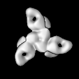
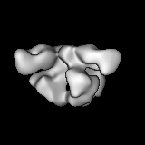
全体 : Complex containing 3 copies of N123-VRC34.01 anti-HIV Fab bound t...
全体
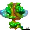
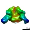
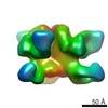
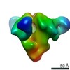
名称: Complex containing 3 copies of N123-VRC34.01 anti-HIV Fab bound to a trimer of HIV-1 Env B505 SOSIP.664
要素
複合体: Complex containing 3 copies of N123-VRC34.01 anti-HIV Fab bound to a trimer of HIV-1 Env B505 SOSIP.664
複合体: HIV-1 Env BG505 SOSIP.664
複合体: Anti-HIV N123-VRC34.01 antibody fragment antigen binding
-
超分子 #1: Complex containing 3 copies of N123-VRC34.01 anti-HIV Fab bound t...
超分子
名称: Complex containing 3 copies of N123-VRC34.01 anti-HIV Fab bound to a trimer of HIV-1 Env B505 SOSIP.664 タイプ: complex / ID: 1 / 親要素: 0
分子量
理論値: 570 KDa
-
超分子 #2: HIV-1 Env BG505 SOSIP.664
超分子
名称: HIV-1 Env BG505 SOSIP.664 / タイプ: complex / ID: 2 / 親要素: 1 詳細: Soluble and stabilized HIV-1 Env trimer from strain BG505. Engineered disulfide between A501C and T605C. I559P mutation to stabilize in pre-fusion state. Addition of N332 to restore ...詳細: Soluble and stabilized HIV-1 Env trimer from strain BG505. Engineered disulfide between A501C and T605C. I559P mutation to stabilize in pre-fusion state. Addition of N332 to restore glycosylation site for purification and antigenic properties. Truncation after D664 to increase solubility. Formed with three gp140 subunits.
由来(天然)
生物種: Human immunodeficiency virus 1 (ヒト免疫不全ウイルス) 株: BG505
組換発現
生物種: Homo sapiens (ヒト) / 組換細胞: HEK293F / 組換プラスミド: pPPI4
-
超分子 #3: Anti-HIV N123-VRC34.01 antibody fragment antigen binding
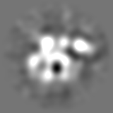
タイプ: PROJECTION MATCHING / ソフトウェア - 名称: SPARX / ソフトウェア - 詳細: sxali3d.py 詳細: Sparx sxali3d.py was used to refine particles against an initial model generated by EMAN2.
 ムービー
ムービー コントローラー


 データを開く
 基本情報
基本情報 マップデータ
 試料
試料 機能・相同性情報
 Human immunodeficiency virus 1 (ヒト免疫不全ウイルス) /
  Homo sapiens (ヒト)
 データ登録者
 引用
引用
 構造の表示
構造の表示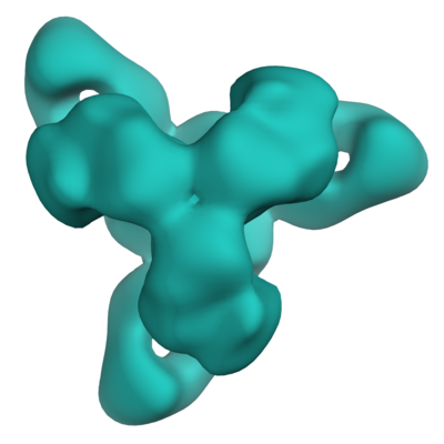
 ダウンロードとリンク
 emd_8125.png
 http://ftp.pdbj.org/pub/emdb/structures/EMD-8125


 Z (Sec.)
 Y (Row.)
 X (Col.)


 試料の構成要素
 解析
解析 電子顕微鏡法
 FIELD EMISSION GUN