[English] 日本語
Yorodumi
- EMDB-71090: Nanodisc-embedded human TF/FVIIa/XK1 in complex with 10H10 Fab (n... -

+
Open data


ID or keywords:

Loading...

-
Basic information

Entry
Database: EMDB / ID: EMD-71090
TitleNanodisc-embedded human TF/FVIIa/XK1 in complex with 10H10 Fab (nanodisc-subtracted)
Map dataCryoEM structure of nanodisc-embedded human blood clotting factor, fVIIa/TF/XK1 in complex with 10H10 Fab.
Sample
  • Complex: Complex of factor VIIa, XK1 and the Fab fragment of antibody 10H10, all bound to tissue factor embedded in a nanodisc membrane bilayer.
    • Complex: Human 10H10 antibody Fab
      • Protein or peptide: x 2 types
    • Complex: XK1 chimeric protein
      • Protein or peptide: x 2 types
    • Complex: Human factor VIIa (FVIIa)
      • Protein or peptide: x 2 types
    • Complex: Human MBP-tagged tissue factor
      • Protein or peptide: x 1 types
  • Ligand: x 4 types
Keywordscomplex / nanodisc / blood clotting
Function / homology
Function and homology information


activation of plasma proteins involved in acute inflammatory response / activation of blood coagulation via clotting cascade / coagulation factor VIIa / response to Thyroid stimulating hormone / response to astaxanthin / response to thyrotropin-releasing hormone / response to 2,3,7,8-tetrachlorodibenzodioxine / response to carbon dioxide / response to genistein / serine-type peptidase complex ...activation of plasma proteins involved in acute inflammatory response / activation of blood coagulation via clotting cascade / coagulation factor VIIa / response to Thyroid stimulating hormone / response to astaxanthin / response to thyrotropin-releasing hormone / response to 2,3,7,8-tetrachlorodibenzodioxine / response to carbon dioxide / response to genistein / serine-type peptidase complex / response to vitamin K / coagulation factor Xa / positive regulation of platelet-derived growth factor receptor signaling pathway / positive regulation of leukocyte chemotaxis / response to thyroxine / NGF-stimulated transcription / Defective factor IX causes thrombophilia / Defective cofactor function of FVIIIa variant / Defective F9 variant does not activate FX / response to cholesterol / cytokine receptor activity / response to growth hormone / positive regulation of positive chemotaxis / Extrinsic Pathway of Fibrin Clot Formation / endopeptidase inhibitor activity / cellular response to steroid hormone stimulus / positive regulation of endothelial cell apoptotic process / negative regulation of blood coagulation / carbohydrate transmembrane transporter activity / positive regulation of blood coagulation / animal organ regeneration / maltose binding / positive regulation of TOR signaling / maltose transport / maltodextrin transmembrane transport / Transport of gamma-carboxylated protein precursors from the endoplasmic reticulum to the Golgi apparatus / Gamma-carboxylation of protein precursors / Common Pathway of Fibrin Clot Formation / Removal of aminoterminal propeptides from gamma-carboxylated proteins / side of membrane / ATP-binding cassette (ABC) transporter complex, substrate-binding subunit-containing / Intrinsic Pathway of Fibrin Clot Formation / positive regulation of endothelial cell proliferation / serine-type peptidase activity / BMAL1:CLOCK,NPAS2 activates circadian expression / positive regulation of interleukin-8 production / serine-type endopeptidase inhibitor activity / circadian rhythm / phospholipid binding / caveola / protein processing / Golgi lumen / response to estrogen / cytokine-mediated signaling pathway / positive regulation of angiogenesis / blood coagulation / response to estradiol / : / outer membrane-bounded periplasmic space / protease binding / vesicle / response to hypoxia / positive regulation of cell migration / endoplasmic reticulum lumen / signaling receptor binding / serine-type endopeptidase activity / external side of plasma membrane / calcium ion binding / positive regulation of gene expression / cell surface / endoplasmic reticulum / proteolysis / extracellular space / extracellular region / membrane / plasma membrane
Similarity search - Function
Tissue factor pathway inhibitor-like / Tissue factor / Tissue factor, conserved site / Tissue factor signature. / Interferon/interleukin receptor domain / Interferon-alpha/beta receptor, fibronectin type III / : / Tissue factor / : / Peptidase S1A, coagulation factor VII/IX/X/C/Z ...Tissue factor pathway inhibitor-like / Tissue factor / Tissue factor, conserved site / Tissue factor signature. / Interferon/interleukin receptor domain / Interferon-alpha/beta receptor, fibronectin type III / : / Tissue factor / : / Peptidase S1A, coagulation factor VII/IX/X/C/Z / : / Coagulation factor-like, Gla domain superfamily / Proteinase inhibitor I2, Kunitz, conserved site / Pancreatic trypsin inhibitor (Kunitz) family signature. / BPTI/Kunitz family of serine protease inhibitors. / Pancreatic trypsin inhibitor Kunitz domain / Kunitz/Bovine pancreatic trypsin inhibitor domain / Pancreatic trypsin inhibitor (Kunitz) family profile. / Pancreatic trypsin inhibitor Kunitz domain superfamily / Coagulation Factor Xa inhibitory site / EGF-like domain / Maltose/Cyclodextrin ABC transporter, substrate-binding protein / EGF-type aspartate/asparagine hydroxylation site / EGF-like calcium-binding, conserved site / Calcium-binding EGF-like domain signature. / Solute-binding family 1, conserved site / Bacterial extracellular solute-binding proteins, family 1 signature. / Aspartic acid and asparagine hydroxylation site. / EGF-like calcium-binding domain / Calcium-binding EGF-like domain / Vitamin K-dependent carboxylation/gamma-carboxyglutamic (GLA) domain / Gamma-carboxyglutamic acid-rich (GLA) domain / Gamma-carboxyglutamic acid-rich (GLA) domain superfamily / Vitamin K-dependent carboxylation domain. / Gla domain profile. / Domain containing Gla (gamma-carboxyglutamate) residues. / Bacterial extracellular solute-binding protein / Bacterial extracellular solute-binding protein / Epidermal growth factor-like domain. / EGF-like domain profile. / EGF-like domain signature 1. / EGF-like domain signature 2. / EGF-like domain / Fibronectin type III / Fibronectin type III superfamily / Serine proteases, trypsin family, histidine active site / Serine proteases, trypsin family, serine active site / Serine proteases, trypsin family, histidine active site. / Peptidase S1A, chymotrypsin family / Serine proteases, trypsin family, serine active site. / Serine proteases, trypsin domain profile. / Trypsin-like serine protease / Serine proteases, trypsin domain / Trypsin / Peptidase S1, PA clan, chymotrypsin-like fold / Immunoglobulin-like fold / Peptidase S1, PA clan
Similarity search - Domain/homology
Coagulation factor X / Coagulation factor VII / Maltose/maltodextrin-binding periplasmic protein / Tissue factor pathway inhibitor / Tissue factor
Similarity search - Component
Biological speciesMus musculus (house mouse) / Homo sapiens (human) / Escherichia coli (E. coli)
Methodsingle particle reconstruction / cryo EM / Resolution: 3.7 Å
AuthorsPhotenhauer AL / Sedzro JC / Ohi MD / Morrissey JH
Funding support United States, 5 items
OrganizationGrant numberCountry
National Institutes of Health/National Heart, Lung, and Blood Institute (NIH/NHLBI)R35 HL135823 United States
National Institutes of Health/National Heart, Lung, and Blood Institute (NIH/NHLBI)R35 HL171334 United States
National Institutes of Health/Office of the DirectorS10OD030275 United States
National Institutes of Health/National Institute of General Medical Sciences (NIH/NIGMS)R24 GM145965 United States
American Heart AssociationN028347 United States
Citation
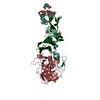
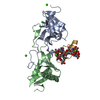
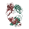
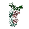
Journal: Blood / Year: 2025
Title: Cryo-EM structure of the tissue factor/factor VIIa complex with a factor X mimetic reveals a novel allosteric mechanism.
Authors: Josepha C Sedzro / Amanda L Photenhauer / Fabienne Birkle / Katarina Meze / Alex Mortenson / Cade Duckworth / Po-Chao Wen / Sarah Kearns / Michael A Cianfrocco / Emad Tajkhorshid / Melanie D ...Authors: Josepha C Sedzro / Amanda L Photenhauer / Fabienne Birkle / Katarina Meze / Alex Mortenson / Cade Duckworth / Po-Chao Wen / Sarah Kearns / Michael A Cianfrocco / Emad Tajkhorshid / Melanie D Ohi / James H Morrissey /
Abstract: Blood clotting is triggered in hemostasis and thrombosis when the membrane-bound tissue factor (TF)/factor VIIa (FVIIa) complex activates factor X (FX). There are no structures of TF/FVIIa on ...Blood clotting is triggered in hemostasis and thrombosis when the membrane-bound tissue factor (TF)/factor VIIa (FVIIa) complex activates factor X (FX). There are no structures of TF/FVIIa on membranes, with or without FX. Using cryo-EM to address this gap, we assembled TF/FVIIa complexes on nanoscale membrane bilayers (nanodiscs), bound to XK1 and an antibody fragment. XK1 is a FX mimetic whose protease domain is replaced by the first Kunitz-type (K1) domain of tissue factor pathway inhibitor, while 10H10 is a non-inhibitory, anti-TF antibody. We determined a cryo-EM structure of a TF/FVIIa/XK1/10H10/nanodisc complex with a resolution of 3.7 Å, allowing us to model all the protein backbones. TF/FVIIa extends perpendicularly from the membrane, interacting with a "handle shaped" XK1 at two locations: the K1 domain docks into FVIIa's active site, while the γ-carboxyglutamate-rich (GLA) domain binds to TF's substrate-binding exosite. The FX and FVIIa GLA domains also contact each other and the membrane surface. Except for a minor contact between the first epidermal growth factor (EGF)-like domain of XK1 and TF, the rest of the FX light chain does not interact with TF/FVIIa. The structure reveals a previously unrecognized, membrane-dependent allosteric activation mechanism between FVIIa and TF where a serine-rich loop in TF that partially obscures the TF exosite must undergo a shift to allow access of the FX GLA domain to its final binding location on the membrane-bound TF/FVIIa complex. This mechanism also provides a novel explanation for the otherwise puzzling phenomenon of TF encryption/decryption on cell surfaces.
History
DepositionJun 7, 2025-
Header (metadata) releaseAug 27, 2025-
Map releaseAug 27, 2025-
UpdateAug 27, 2025-
Current statusAug 27, 2025Processing site: RCSB / Status: Released

-
Structure visualization

Downloads & links

-
Map

FileDownload / File: emd_71090.map.gz / Format: CCP4 / Size: 282.6 MB / Type: IMAGE STORED AS FLOATING POINT NUMBER (4 BYTES)
AnnotationCryoEM structure of nanodisc-embedded human blood clotting factor, fVIIa/TF/XK1 in complex with 10H10 Fab.
Voxel sizeX=Y=Z: 0.834 Å
Density
Contour LevelBy AUTHOR: 0.04
Minimum - Maximum-0.23646995 - 0.4661354
Average (Standard dev.)-0.000073506766 (±0.009938195)
SymmetrySpace group: 1
Details

EMDB XML:

Map geometry
Axis orderXYZ
Origin000
Dimensions420420420
Spacing420420420
CellA=B=C: 350.28 Å
α=β=γ: 90.0 °

-
Supplemental data

-
Sample components

+
Entire : Complex of factor VIIa, XK1 and the Fab fragment of antibody 10H1...

EntireName: Complex of factor VIIa, XK1 and the Fab fragment of antibody 10H10, all bound to tissue factor embedded in a nanodisc membrane bilayer.
Components
  • Complex: Complex of factor VIIa, XK1 and the Fab fragment of antibody 10H10, all bound to tissue factor embedded in a nanodisc membrane bilayer.
    • Complex: Human 10H10 antibody Fab
      • Protein or peptide: Human 10H10 antibody Fab heavy chain
      • Protein or peptide: Human 10H10 antibody Fab light chain
    • Complex: XK1 chimeric protein
      • Protein or peptide: Factor X light chain
      • Protein or peptide: Tissue factor pathway inhibitor
    • Complex: Human factor VIIa (FVIIa)
      • Protein or peptide: Factor VII light chain
      • Protein or peptide: Coagulation factor VII Heavy Chain
    • Complex: Human MBP-tagged tissue factor
      • Protein or peptide: Tissue factor,Maltose/maltodextrin-binding periplasmic protein
  • Ligand: MAGNESIUM ION
  • Ligand: CALCIUM ION
  • Ligand: beta-D-glucopyranose
  • Ligand: alpha-L-fucopyranose

+
Supramolecule #1: Complex of factor VIIa, XK1 and the Fab fragment of antibody 10H1...

SupramoleculeName: Complex of factor VIIa, XK1 and the Fab fragment of antibody 10H10, all bound to tissue factor embedded in a nanodisc membrane bilayer.
type: complex / ID: 1 / Parent: 0 / Macromolecule list: #1-#7 / Details: Map with nanodisc subtracted.

+
Supramolecule #2: Human 10H10 antibody Fab

SupramoleculeName: Human 10H10 antibody Fab / type: complex / ID: 2 / Parent: 1 / Macromolecule list: #6-#7
Source (natural)Organism: Mus musculus (house mouse)

+
Supramolecule #3: XK1 chimeric protein

SupramoleculeName: XK1 chimeric protein / type: complex / ID: 3 / Parent: 1 / Macromolecule list: #2, #5
Source (natural)Organism: Homo sapiens (human)

+
Supramolecule #4: Human factor VIIa (FVIIa)

SupramoleculeName: Human factor VIIa (FVIIa) / type: complex / ID: 4 / Parent: 1 / Macromolecule list: #1, #3
Source (natural)Organism: Homo sapiens (human)

+
Supramolecule #5: Human MBP-tagged tissue factor

SupramoleculeName: Human MBP-tagged tissue factor / type: complex / ID: 5 / Parent: 1 / Macromolecule list: #4
Source (natural)Organism: Homo sapiens (human)

+
Macromolecule #1: Factor VII light chain

MacromoleculeName: Factor VII light chain / type: protein_or_peptide / ID: 1 / Number of copies: 1 / Enantiomer: LEVO
Source (natural)Organism: Homo sapiens (human)
Molecular weightTheoretical: 16.359772 KDa
Recombinant expressionOrganism: Homo sapiens (human)
SequenceString:
ANAFL(CGU)(CGU)LRP GSL(CGU)R(CGU)CK(CGU)(CGU) QCSF(CGU)(CGU)AR(CGU)I FKDA(CGU)RTKLF WISYSDGDQ CASSPCQNGG SCKDQLQSYI CFCLPAFEGR NCETHKDDQL ICVNENGGCE QYCSDHTGTK RSCRCHEGYS L LADGVSCT PTVEYPCGKI PILE

UniProtKB: Coagulation factor VII

+
Macromolecule #2: Factor X light chain

MacromoleculeName: Factor X light chain / type: protein_or_peptide / ID: 2 / Number of copies: 1 / Enantiomer: LEVO
Source (natural)Organism: Homo sapiens (human)
Molecular weightTheoretical: 15.309423 KDa
Recombinant expressionOrganism: Homo sapiens (human)
SequenceString:
ANSFL(CGU)(CGU)MKK GHL(CGU)R(CGU)CM(CGU)(CGU) TCSY(CGU)(CGU)AR(CGU)V F(CGU)DSDKTN (CGU) FWNKYKDGDQ CETSPCQNQG KCKDGLGEYT CTCLEGFEGK NCELFTRKLC SLDNGDCDQF CHEEQNSVVC SCARG YTLA DNGKACIPTG PYP

UniProtKB: Coagulation factor X

+
Macromolecule #3: Coagulation factor VII Heavy Chain

MacromoleculeName: Coagulation factor VII Heavy Chain / type: protein_or_peptide / ID: 3 / Number of copies: 1 / Enantiomer: LEVO / EC number: coagulation factor VIIa
Source (natural)Organism: Homo sapiens (human)
Molecular weightTheoretical: 28.103256 KDa
Recombinant expressionOrganism: Homo sapiens (human)
SequenceString: IVGGKVCPKG ECPWQVLLLV NGAQLCGGTL INTIWVVSAA HCFDKIKNWR NLIAVLGEHD LSEHDGDEQS RRVAQVIIPS TYVPGTTNH DIALLRLHQP VVLTDHVVPL CLPERTFSER TLAFVRFSLV SGWGQLLDRG ATALELMVLN VPRLMTQDCL Q QSRKVGDS ...String:
IVGGKVCPKG ECPWQVLLLV NGAQLCGGTL INTIWVVSAA HCFDKIKNWR NLIAVLGEHD LSEHDGDEQS RRVAQVIIPS TYVPGTTNH DIALLRLHQP VVLTDHVVPL CLPERTFSER TLAFVRFSLV SGWGQLLDRG ATALELMVLN VPRLMTQDCL Q QSRKVGDS PNITEYMFCA GYSDGSKDSC KGDSGGPHAT HYRGTWYLTG IVSWGQGCAT VGHFGVYTRV SQYIEWLQKL MR SEPRPGV LLRAPFP

UniProtKB: Coagulation factor VII

+
Macromolecule #4: Tissue factor,Maltose/maltodextrin-binding periplasmic protein

MacromoleculeName: Tissue factor,Maltose/maltodextrin-binding periplasmic protein
type: protein_or_peptide / ID: 4 / Number of copies: 1 / Enantiomer: LEVO
Source (natural)Organism: Escherichia coli (E. coli)
Molecular weightTheoretical: 67.828523 KDa
Recombinant expressionOrganism: Escherichia coli BL21(DE3) (bacteria)
SequenceString: TVAAYNLTWK STNFKTILEW EPKPVNQVYT VQISTKSGDW KSKCFYTTDT ECDLTDEIVK DVKQTYLARV FSYPAGNVES TGSAGEPLY ENSPEFTPYL ETNLGQPTIQ SFEQVGTKVN VTVEDERTLV RRNNTFLSLR DVFGKDLIYT LYYWKSSSSG K KTAKTNTN ...String:
TVAAYNLTWK STNFKTILEW EPKPVNQVYT VQISTKSGDW KSKCFYTTDT ECDLTDEIVK DVKQTYLARV FSYPAGNVES TGSAGEPLY ENSPEFTPYL ETNLGQPTIQ SFEQVGTKVN VTVEDERTLV RRNNTFLSLR DVFGKDLIYT LYYWKSSSSG K KTAKTNTN EFLIDVDKGE NYCFSVQAVI PSRTVNRKST DSPVECMGQE KGEFREIFYI IGAVVFVVII LVIILAISLH KG SGSGMKI EEGKLVIWIN GDKGYNGLAE VGKKFEKDTG IKVTVEHPDK LEEKFPQVAA TGDGPDIIFW AHDRFGGYAQ SGL LAEITP DKAFQDKLYP FTWDAVRYNG KLIAYPIAVE ALSLIYNKDL LPNPPKTWEE IPALDKELKA KGKSALMFNL QEPY FTWPL IAADGGYAFK YENGKYDIKD VGVDNAGAKA GLTFLVDLIK NKHMNADTDY SIAEAAFNKG ETAMTINGPW AWSNI DTSK VNYGVTVLPT FKGQPSKPFV GVLSAGINAA SPNKELAKEF LENYLLTDEG LEAVNKDKPL GAVALKSYEE ELAKDP RIA ATMENAQKGE IMPNIPQMSA FWYAVRTAVI NAASGRQTVD EALKDAQT

UniProtKB: Tissue factor, Maltose/maltodextrin-binding periplasmic protein

+
Macromolecule #5: Tissue factor pathway inhibitor

MacromoleculeName: Tissue factor pathway inhibitor / type: protein_or_peptide / ID: 5 / Number of copies: 1 / Enantiomer: LEVO
Source (natural)Organism: Homo sapiens (human)
Molecular weightTheoretical: 7.979198 KDa
Recombinant expressionOrganism: Homo sapiens (human)
SequenceString:
TDTELPPLKL MHSFCAFKAD DGPCKAIMKR FFFNIFTRQC EEFIYGGCEG NQNRFESLEE CKKMCTRD

UniProtKB: Tissue factor pathway inhibitor

+
Macromolecule #6: Human 10H10 antibody Fab heavy chain

MacromoleculeName: Human 10H10 antibody Fab heavy chain / type: protein_or_peptide / ID: 6 / Number of copies: 1 / Enantiomer: LEVO
Source (natural)Organism: Mus musculus (house mouse) / Strain: BALB/c
Molecular weightTheoretical: 23.569252 KDa
SequenceString: QVHLQQSGAE LMKPGASVKI SCKASGYTFI TYWIEWVKQR PGHGLEWIGD ILPGSGSTNY NENFKGKATF TADSSSNTAY MQLSSLTSE DSAVYYCARS GYYGNSGFAY WGQGTLVTVS AAKTTPPSVY PLAPGSAAQT NSMVTLGCLV KGYFPEPVTV T WNSGSLSS ...String:
QVHLQQSGAE LMKPGASVKI SCKASGYTFI TYWIEWVKQR PGHGLEWIGD ILPGSGSTNY NENFKGKATF TADSSSNTAY MQLSSLTSE DSAVYYCARS GYYGNSGFAY WGQGTLVTVS AAKTTPPSVY PLAPGSAAQT NSMVTLGCLV KGYFPEPVTV T WNSGSLSS GVHTFPAVLQ SDLYTLSSSV TVPSSTWPSE TVTCNVAHPA SSTKVDKKIV PRD

+
Macromolecule #7: Human 10H10 antibody Fab light chain

MacromoleculeName: Human 10H10 antibody Fab light chain / type: protein_or_peptide / ID: 7 / Number of copies: 1 / Enantiomer: LEVO
Source (natural)Organism: Mus musculus (house mouse) / Strain: BALB/c
Molecular weightTheoretical: 24.196709 KDa
SequenceString: DIVMTQSPSS LTVTAGEKVT MSCKSSQSLL SSGNQKNYLT WYQQIPGQPP KLLIYWASTR ESGVPDRFTG SGSGTDFTLT INSVQAEDL AVYYCQNDYT YPLTFGAGTK LELKRADAAP TVSIFPPSSE QLTSGGASVV CFLNNFYPKD INVKWKIDGS E RQNGVLNS ...String:
DIVMTQSPSS LTVTAGEKVT MSCKSSQSLL SSGNQKNYLT WYQQIPGQPP KLLIYWASTR ESGVPDRFTG SGSGTDFTLT INSVQAEDL AVYYCQNDYT YPLTFGAGTK LELKRADAAP TVSIFPPSSE QLTSGGASVV CFLNNFYPKD INVKWKIDGS E RQNGVLNS ATDQDSKDST YSMSSTLTLT KDEYERHNSY TCEATHKTST SPIVKSFNRN EC

+
Macromolecule #8: MAGNESIUM ION

MacromoleculeName: MAGNESIUM ION / type: ligand / ID: 8 / Number of copies: 4 / Formula: MG
Molecular weightTheoretical: 24.305 Da

+
Macromolecule #9: CALCIUM ION

MacromoleculeName: CALCIUM ION / type: ligand / ID: 9 / Number of copies: 12 / Formula: CA
Molecular weightTheoretical: 40.078 Da

+
Macromolecule #10: beta-D-glucopyranose

MacromoleculeName: beta-D-glucopyranose / type: ligand / ID: 10 / Number of copies: 1 / Formula: BGC
Molecular weightTheoretical: 180.156 Da
Chemical component information

ChemComp-BGC:
beta-D-glucopyranose

+
Macromolecule #11: alpha-L-fucopyranose

MacromoleculeName: alpha-L-fucopyranose / type: ligand / ID: 11 / Number of copies: 1 / Formula: FUC
Molecular weightTheoretical: 164.156 Da
Chemical component information

ChemComp-FUC:
alpha-L-fucopyranose

-
Experimental details

-
Structure determination

Methodcryo EM
Processingsingle particle reconstruction
Aggregation stateparticle

-
Sample preparation

BufferpH: 7.4
GridModel: Quantifoil R2/2 / Material: COPPER / Mesh: 200 / Pretreatment - Type: GLOW DISCHARGE / Pretreatment - Time: 30 sec.
VitrificationCryogen name: ETHANE / Chamber humidity: 100 % / Chamber temperature: 277 K / Instrument: FEI VITROBOT MARK IV

-
Electron microscopy

MicroscopeTFS KRIOS
Specialist opticsEnergy filter - Slit width: 20 eV
Image recordingFilm or detector model: GATAN K3 (6k x 4k) / Number grids imaged: 1 / Number real images: 7132 / Average electron dose: 50.0 e/Å2
Electron beamAcceleration voltage: 300 kV / Electron source: FIELD EMISSION GUN
Electron opticsIllumination mode: FLOOD BEAM / Imaging mode: BRIGHT FIELD / Nominal defocus max: 2.5 µm / Nominal defocus min: 1.0 µm / Nominal magnification: 105000
Experimental equipment
Model: Titan Krios / Image courtesy: FEI Company

+
Image processing

Particle selectionNumber selected: 2862371
CTF correctionType: NONE
Startup modelType of model: NONE
Final reconstructionApplied symmetry - Point group: C1 (asymmetric) / Resolution.type: BY AUTHOR / Resolution: 3.7 Å / Resolution method: FSC 0.143 CUT-OFF / Software - Name: cryoSPARC / Number images used: 20107
Initial angle assignmentType: MAXIMUM LIKELIHOOD
Final angle assignmentType: MAXIMUM LIKELIHOOD

-
Atomic model buiding 1

Initial model
PDB IDChain

chain_id: L, residue_range: 46-142, source_name: PDB, initial_model_type: experimental model

chain_id: H, residue_range: 16-257, source_name: PDB, initial_model_type: experimental model

chain_id: T, residue_range: 6-80, source_name: PDB, initial_model_type: experimental model

chain_id: U, residue_range: 91-210, source_name: PDB, initial_model_type: experimental model

chain_id: L, residue_range: 1-46, source_name: PDB, initial_model_type: experimental model

chain_id: G, residue_range: 401-444, source_name: PDB, initial_model_type: experimental model

chain_id: L, residue_range: 49-139, source_name: PDB, initial_model_type: experimental model

chain_id: A, residue_range: 12-79, source_name: PDB, initial_model_type: experimental model

chain_id: H, residue_range: 112-214, source_name: PDB, initial_model_type: experimental model

chain_id: L, residue_range: 107-214, source_name: PDB, initial_model_type: experimental model

chain_id: H, residue_range: 1-119, source_name: PDB, initial_model_type: experimental model

chain_id: L, residue_range: 1-113, source_name: PDB, initial_model_type: experimental model
DetailsCrystal structures were rigid-body fit into the density map and model optimization was then carried out with Phenix real-space refine. Manual refinement in Coot was performed to ensure that the backbone traces followed the density in regions where the map was high enough resolution to trace the backbone. Secondary structure restraints were used to ensure that alpha-helices and beta-sheets did not deviate far from their expected geometry. A final check of MolProbity and cross correlation was done to ensure model quality.
RefinementSpace: REAL / Protocol: FLEXIBLE FIT / Target criteria: cross-correlation
Output model

PDB-9p0x:
Nanodisc-embedded human TF/FVIIa/XK1 in complex with 10H10 Fab (nanodisc-subtracted)

+
About Yorodumi

-
News

-
Feb 9, 2022. New format data for meta-information of EMDB entries

New format data for meta-information of EMDB entries

  • Version 3 of the EMDB header file is now the official format.
  • The previous official version 1.9 will be removed from the archive.

Related info.:EMDB header

External links:wwPDB to switch to version 3 of the EMDB data model

-
Aug 12, 2020. Covid-19 info

Covid-19 info

URL: https://pdbj.org/emnavi/covid19.php

New page: Covid-19 featured information page in EM Navigator.

Related info.:Covid-19 info / Mar 5, 2020. Novel coronavirus structure data

+
Mar 5, 2020. Novel coronavirus structure data

Novel coronavirus structure data

Related info.:Yorodumi Speices / Aug 12, 2020. Covid-19 info

External links:COVID-19 featured content - PDBj / Molecule of the Month (242):Coronavirus Proteases

+
Jan 31, 2019. EMDB accession codes are about to change! (news from PDBe EMDB page)

EMDB accession codes are about to change! (news from PDBe EMDB page)

  • The allocation of 4 digits for EMDB accession codes will soon come to an end. Whilst these codes will remain in use, new EMDB accession codes will include an additional digit and will expand incrementally as the available range of codes is exhausted. The current 4-digit format prefixed with “EMD-” (i.e. EMD-XXXX) will advance to a 5-digit format (i.e. EMD-XXXXX), and so on. It is currently estimated that the 4-digit codes will be depleted around Spring 2019, at which point the 5-digit format will come into force.
  • The EM Navigator/Yorodumi systems omit the EMD- prefix.

Related info.:Q: What is EMD? / ID/Accession-code notation in Yorodumi/EM Navigator

External links:EMDB Accession Codes are Changing Soon! / Contact to PDBj

+
Jul 12, 2017. Major update of PDB

Major update of PDB

  • wwPDB released updated PDB data conforming to the new PDBx/mmCIF dictionary.
  • This is a major update changing the version number from 4 to 5, and with Remediation, in which all the entries are updated.
  • In this update, many items about electron microscopy experimental information are reorganized (e.g. em_software).
  • Now, EM Navigator and Yorodumi are based on the updated data.

External links:wwPDB Remediation / Enriched Model Files Conforming to OneDep Data Standards Now Available in the PDB FTP Archive

-
Yorodumi

Thousand views of thousand structures

  • Yorodumi is a browser for structure data from EMDB, PDB, SASBDB, etc.
  • This page is also the successor to EM Navigator detail page, and also detail information page/front-end page for Omokage search.
  • The word "yorodu" (or yorozu) is an old Japanese word meaning "ten thousand". "mi" (miru) is to see.

Related info.:EMDB / PDB / SASBDB / Comparison of 3 databanks / Yorodumi Search / Aug 31, 2016. New EM Navigator & Yorodumi / Yorodumi Papers / Jmol/JSmol / Function and homology information / Changes in new EM Navigator and Yorodumi

Read more