Movie
Movie Controller

[English] 日本語
 Yorodumi
- PDB-9p0x: Nanodisc-embedded human TF/FVIIa/XK1 in complex with 10H10 Fab (n... -
+ Open data

- Basic information
| Entry | Database: PDB / ID: 9p0x | ||||||||||||||||||||||||
|---|---|---|---|---|---|---|---|---|---|---|---|---|---|---|---|---|---|---|---|---|---|---|---|---|---|
| Title | Nanodisc-embedded human TF/FVIIa/XK1 in complex with 10H10 Fab (nanodisc-subtracted) | ||||||||||||||||||||||||
 Components Components |
| ||||||||||||||||||||||||
 Keywords Keywords | BLOOD CLOTTING / complex / nanodisc | ||||||||||||||||||||||||
| Function / homology |  Function and homology information Function and homology informationactivation of plasma proteins involved in acute inflammatory response / activation of blood coagulation via clotting cascade / coagulation factor VIIa / response to Thyroid stimulating hormone / response to astaxanthin / response to thyrotropin-releasing hormone / response to 2,3,7,8-tetrachlorodibenzodioxine / response to carbon dioxide / serine-type peptidase complex / response to genistein ...activation of plasma proteins involved in acute inflammatory response / activation of blood coagulation via clotting cascade / coagulation factor VIIa / response to Thyroid stimulating hormone / response to astaxanthin / response to thyrotropin-releasing hormone / response to 2,3,7,8-tetrachlorodibenzodioxine / response to carbon dioxide / serine-type peptidase complex / response to genistein / response to vitamin K / coagulation factor Xa / positive regulation of platelet-derived growth factor receptor signaling pathway / positive regulation of leukocyte chemotaxis / response to thyroxine / NGF-stimulated transcription / Defective factor IX causes thrombophilia / Defective cofactor function of FVIIIa variant / Defective F9 variant does not activate FX / response to cholesterol / cytokine receptor activity / positive regulation of positive chemotaxis / : / response to growth hormone / cellular response to steroid hormone stimulus / endopeptidase inhibitor activity / negative regulation of blood coagulation / positive regulation of blood coagulation / animal organ regeneration / positive regulation of endothelial cell apoptotic process / positive regulation of TOR signaling / carbohydrate transmembrane transporter activity / Transport of gamma-carboxylated protein precursors from the endoplasmic reticulum to the Golgi apparatus / maltose binding / : / Gamma-carboxylation of protein precursors / maltose transport / maltodextrin transmembrane transport / Removal of aminoterminal propeptides from gamma-carboxylated proteins / side of membrane / : / ATP-binding cassette (ABC) transporter complex, substrate-binding subunit-containing / positive regulation of endothelial cell proliferation / BMAL1:CLOCK,NPAS2 activates circadian expression / serine-type peptidase activity / positive regulation of interleukin-8 production / serine-type endopeptidase inhibitor activity / circadian rhythm / protein processing / phospholipid binding / caveola / response to estrogen / Golgi lumen / cytokine-mediated signaling pathway / positive regulation of angiogenesis / blood coagulation / response to estradiol / outer membrane-bounded periplasmic space / extracellular matrix / protease binding / vesicle / response to hypoxia / positive regulation of cell migration / endoplasmic reticulum lumen / signaling receptor binding / serine-type endopeptidase activity / external side of plasma membrane / calcium ion binding / positive regulation of gene expression / cell surface / proteolysis / : / extracellular region / membrane / plasma membrane Similarity search - Function | ||||||||||||||||||||||||
| Biological species |  Homo sapiens (human) Homo sapiens (human)  | ||||||||||||||||||||||||
| Method | ELECTRON MICROSCOPY / single particle reconstruction / cryo EM / Resolution: 3.7 Å | ||||||||||||||||||||||||
 Authors Authors | Photenhauer, A.L. / Sedzro, J.C. / Ohi, M.D. / Morrissey, J.H. | ||||||||||||||||||||||||
| Funding support |  United States, 5items United States, 5items
| ||||||||||||||||||||||||
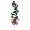
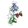
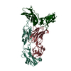
 Citation Citation | Journal: Blood / Year: 2025 Title: Cryo-EM structure of the tissue factor/factor VIIa complex with a factor X mimetic reveals a novel allosteric mechanism. Authors: Josepha C Sedzro / Amanda L Photenhauer / Fabienne Birkle / Katarina Meze / Alex Mortenson / Cade Duckworth / Po-Chao Wen / Sarah Kearns / Michael A Cianfrocco / Emad Tajkhorshid / Melanie D ...Authors: Josepha C Sedzro / Amanda L Photenhauer / Fabienne Birkle / Katarina Meze / Alex Mortenson / Cade Duckworth / Po-Chao Wen / Sarah Kearns / Michael A Cianfrocco / Emad Tajkhorshid / Melanie D Ohi / James H Morrissey / Abstract: Blood clotting is triggered in hemostasis and thrombosis when the membrane-bound tissue factor (TF)/factor VIIa (FVIIa) complex activates factor X (FX). There are no structures of TF/FVIIa on ...Blood clotting is triggered in hemostasis and thrombosis when the membrane-bound tissue factor (TF)/factor VIIa (FVIIa) complex activates factor X (FX). There are no structures of TF/FVIIa on membranes, with or without FX. Using cryoelectron microscopy (cryo-EM) to address this gap, we assembled TF/FVIIa complexes on nanoscale membrane bilayers (nanodiscs), bound to XK1 and an antibody fragment. XK1 is a FX mimetic whose protease domain is replaced by the first Kunitz-type (K1) domain of the TF pathway inhibitor, whereas 10H10 is a noninhibitory, anti-TF antibody. We determined a cryo-EM structure of a TF/FVIIa/XK1/10H10/nanodisc complex with a resolution of 3.7 Å, allowing us to model all the protein backbones. TF/FVIIa extends perpendicularly from the membrane, interacting with a "handle-shaped" XK1 at 2 locations: the K1 domain docks into FVIIa's active site, whereas the γ-carboxyglutamate-rich (GLA) domain binds to the TF substrate-binding exosite. The FX and FVIIa GLA domains also contact each other and the membrane surface. Except for a minor contact between the first epidermal growth factor (EGF)-like domain of XK1 and TF, the rest of the FX light chain does not interact with TF/FVIIa. The structure reveals a previously unrecognized, membrane-dependent allosteric activation mechanism between FVIIa and TF, in which a serine-rich loop in TF that partially obscures the TF exosite must undergo a shift to allow access of the FX GLA domain to its final binding location on the membrane-bound TF/FVIIa complex. This mechanism also provides a novel explanation for the otherwise puzzling phenomenon of TF encryption/decryption on cell surfaces. | ||||||||||||||||||||||||
| History |
|
- Structure visualization
| Structure viewer | Molecule: MolmilJmol/JSmol |
|---|
- Downloads & links
-Download
| PDBx/mmCIF format | 9p0x.cif.gz | 290.1 KB | Display | PDBx/mmCIF format |
|---|---|---|---|---|
| PDB format | pdb9p0x.ent.gz | 188.3 KB | Display | PDB format |
| PDBx/mmJSON format | 9p0x.json.gz | Tree view | PDBx/mmJSON format | |
| Others |  Other downloads Other downloads |
-Validation report
| Arichive directory | https://data.pdbj.org/pub/pdb/validation_reports/p0/9p0xftp://data.pdbj.org/pub/pdb/validation_reports/p0/9p0x | HTTPS FTP |
|---|
-Related structure data
| Related structure data |  71090MC M: map data used to model this data C: citing same article ( |
|---|---|
| Similar structure data |


-Links
 PDBj
PDBj


- Assembly
| Deposited unit | 
|
|---|---|
| 1 |
|
-Components
-Protein , 5 types, 5 molecules VXSTK
| #1: Protein | Mass: 16359.772 Da / Num. of mol.: 1 Source method: isolated from a genetically manipulated source Source: (gene. exp.) Homo sapiens (human) / Gene: F7 / Cell line (production host): HEK293 / Production host: Homo sapiens (human) / References: UniProt: P08709 |
|---|---|
| #2: Protein | Mass: 15309.423 Da / Num. of mol.: 1 Source method: isolated from a genetically manipulated source Source: (gene. exp.) Homo sapiens (human) / Gene: F10 / Plasmid: pcDNA3.1(+) / Cell line (production host): HEK293 / Production host: Homo sapiens (human) / References: UniProt: P00742 |
| #3: Protein | Mass: 28103.256 Da / Num. of mol.: 1 Source method: isolated from a genetically manipulated source Source: (gene. exp.) Homo sapiens (human) / Gene: F7 / Cell line (production host): HEK293 / Production host: Homo sapiens (human) / References: UniProt: P08709, coagulation factor VIIa |
| #4: Protein | Mass: 67828.523 Da / Num. of mol.: 1 Source method: isolated from a genetically manipulated source Source: (gene. exp.) Homo sapiens (human), (gene. exp.) Gene: F3, malE, Z5632, ECs5017 / Plasmid: pET-26b(+) / Production host: |
| #5: Protein | Mass: 7979.198 Da / Num. of mol.: 1 Source method: isolated from a genetically manipulated source Source: (gene. exp.) Homo sapiens (human) / Gene: TFPI, LACI, TFPI1 / Plasmid: pcDNA3.1(+) / Cell line (production host): HEK293 / Production host: Homo sapiens (human) / References: UniProt: P10646 |
-Antibody , 2 types, 2 molecules HL
| #6: Antibody | Mass: 23569.252 Da / Num. of mol.: 1 / Source method: isolated from a natural source / Source: (natural) |
|---|---|
| #7: Antibody | Mass: 24196.709 Da / Num. of mol.: 1 / Source method: isolated from a natural source / Source: (natural) |
-Sugars , 2 types, 2 molecules 

| #10: Sugar | ChemComp-BGC /  Type: D-saccharide, beta linking / Mass: 180.156 Da / Num. of mol.: 1 / Source method: obtained synthetically / Formula: C6H12O6 Type: D-saccharide, beta linking / Mass: 180.156 Da / Num. of mol.: 1 / Source method: obtained synthetically / Formula: C6H12O6 |
|---|---|
| #11: Sugar | ChemComp-FUC /  Type: L-saccharide, alpha linking / Mass: 164.156 Da / Num. of mol.: 1 / Source method: obtained synthetically / Formula: C6H12O5 Type: L-saccharide, alpha linking / Mass: 164.156 Da / Num. of mol.: 1 / Source method: obtained synthetically / Formula: C6H12O5 |
-Non-polymers , 2 types, 16 molecules 

| #8: Chemical | ChemComp-MG /  Mass: 24.305 Da / Num. of mol.: 4 / Source method: obtained synthetically / Formula: Mg Mass: 24.305 Da / Num. of mol.: 4 / Source method: obtained synthetically / Formula: Mg#9: Chemical | ChemComp-CA /  Mass: 40.078 Da / Num. of mol.: 12 / Source method: obtained synthetically / Formula: Ca Mass: 40.078 Da / Num. of mol.: 12 / Source method: obtained synthetically / Formula: Ca |
|---|
-Details
| Has ligand of interest | N |
|---|---|
| Has protein modification | Y |
-Experimental details
-Experiment
| Experiment | Method: ELECTRON MICROSCOPY |
|---|---|
| EM experiment | Aggregation state: PARTICLE / 3D reconstruction method: single particle reconstruction |
- Sample preparation
| Component |
| ||||||||||||||||||||||||||||||||||||||||||
|---|---|---|---|---|---|---|---|---|---|---|---|---|---|---|---|---|---|---|---|---|---|---|---|---|---|---|---|---|---|---|---|---|---|---|---|---|---|---|---|---|---|---|---|
| Source (natural) |
| ||||||||||||||||||||||||||||||||||||||||||
| Source (recombinant) |
| ||||||||||||||||||||||||||||||||||||||||||
| Buffer solution | pH: 7.4 | ||||||||||||||||||||||||||||||||||||||||||
| Specimen | Embedding applied: NO / Shadowing applied: NO / Staining applied: NO / Vitrification applied: YES | ||||||||||||||||||||||||||||||||||||||||||
| Specimen support | Grid material: COPPER / Grid mesh size: 200 divisions/in. / Grid type: Quantifoil R2/2 | ||||||||||||||||||||||||||||||||||||||||||
| Vitrification | Instrument: FEI VITROBOT MARK IV / Cryogen name: ETHANE / Humidity: 100 % / Chamber temperature: 277 K |
- Electron microscopy imaging
| Experimental equipment |  Model: Titan Krios / Image courtesy: FEI Company |
|---|---|
| Microscopy | Model: TFS KRIOS |
| Electron gun | Electron source:  FIELD EMISSION GUN / Accelerating voltage: 300 kV / Illumination mode: FLOOD BEAM FIELD EMISSION GUN / Accelerating voltage: 300 kV / Illumination mode: FLOOD BEAM |
| Electron lens | Mode: BRIGHT FIELD / Nominal magnification: 105000 X / Nominal defocus max: 2500 nm / Nominal defocus min: 1000 nm |
| Image recording | Electron dose: 50 e/Å2 / Film or detector model: GATAN K3 (6k x 4k) / Num. of grids imaged: 1 / Num. of real images: 7132 |
| EM imaging optics | Energyfilter slit width: 20 eV |
- Processing
| EM software |
| ||||||||||||||||||||||||||||||||||||||||||||||||||||||||||||||||||||||||||||||||||||||||||||||||||||||||
|---|---|---|---|---|---|---|---|---|---|---|---|---|---|---|---|---|---|---|---|---|---|---|---|---|---|---|---|---|---|---|---|---|---|---|---|---|---|---|---|---|---|---|---|---|---|---|---|---|---|---|---|---|---|---|---|---|---|---|---|---|---|---|---|---|---|---|---|---|---|---|---|---|---|---|---|---|---|---|---|---|---|---|---|---|---|---|---|---|---|---|---|---|---|---|---|---|---|---|---|---|---|---|---|---|---|
| CTF correction | Type: NONE | ||||||||||||||||||||||||||||||||||||||||||||||||||||||||||||||||||||||||||||||||||||||||||||||||||||||||
| Particle selection | Num. of particles selected: 2862371 | ||||||||||||||||||||||||||||||||||||||||||||||||||||||||||||||||||||||||||||||||||||||||||||||||||||||||
| Symmetry | Point symmetry: C1 (asymmetric) | ||||||||||||||||||||||||||||||||||||||||||||||||||||||||||||||||||||||||||||||||||||||||||||||||||||||||
| 3D reconstruction | Resolution: 3.7 Å / Resolution method: FSC 0.143 CUT-OFF / Num. of particles: 20107 / Symmetry type: POINT | ||||||||||||||||||||||||||||||||||||||||||||||||||||||||||||||||||||||||||||||||||||||||||||||||||||||||
| Atomic model building | Protocol: FLEXIBLE FIT / Space: REAL / Target criteria: cross-correlation Details: Crystal structures were rigid-body fit into the density map and model optimization was then carried out with Phenix real-space refine. Manual refinement in Coot was performed to ensure that ...Details: Crystal structures were rigid-body fit into the density map and model optimization was then carried out with Phenix real-space refine. Manual refinement in Coot was performed to ensure that the backbone traces followed the density in regions where the map was high enough resolution to trace the backbone. Secondary structure restraints were used to ensure that alpha-helices and beta-sheets did not deviate far from their expected geometry. A final check of MolProbity and cross correlation was done to ensure model quality. | ||||||||||||||||||||||||||||||||||||||||||||||||||||||||||||||||||||||||||||||||||||||||||||||||||||||||
| Atomic model building | 3D fitting-ID: 1 / Source name: PDB / Type: experimental model
| ||||||||||||||||||||||||||||||||||||||||||||||||||||||||||||||||||||||||||||||||||||||||||||||||||||||||
| Refinement | Cross valid method: NONE Stereochemistry target values: GeoStd + Monomer Library + CDL v1.2 | ||||||||||||||||||||||||||||||||||||||||||||||||||||||||||||||||||||||||||||||||||||||||||||||||||||||||
| Displacement parameters | Biso mean: 95.04 Å2 | ||||||||||||||||||||||||||||||||||||||||||||||||||||||||||||||||||||||||||||||||||||||||||||||||||||||||
| Refine LS restraints |
|