 Movie
Movie Controller

[English] 日本語
 Yorodumi
- EMDB-70925: Heteromeric GluA1/A2 in the desensitized state, composite map of ... -
+ Open data

- Basic information
| Entry |  | ||||||||||||||||||||||||||||||
|---|---|---|---|---|---|---|---|---|---|---|---|---|---|---|---|---|---|---|---|---|---|---|---|---|---|---|---|---|---|---|---|
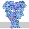
| Title | Heteromeric GluA1/A2 in the desensitized state, composite map of ATD-LBD-TMD | ||||||||||||||||||||||||||||||
 Map data Map data | A1A2 desensitized state (composite map) | ||||||||||||||||||||||||||||||
 Sample Sample |
| ||||||||||||||||||||||||||||||
 Keywords Keywords | GluA1A2 heterotetramer quisqualate iGluR / MEMBRANE PROTEIN | ||||||||||||||||||||||||||||||
| Function / homology |  Function and homology information Function and homology informationCargo concentration in the ER / cellular response to ammonium ion / axonal spine / COPII-mediated vesicle transport / positive regulation of locomotion involved in locomotory behavior / positive regulation of membrane potential / response to sucrose / regulation of monoatomic ion transmembrane transport / myosin V binding / cellular response to L-glutamate ...Cargo concentration in the ER / cellular response to ammonium ion / axonal spine / COPII-mediated vesicle transport / positive regulation of locomotion involved in locomotory behavior / positive regulation of membrane potential / response to sucrose / regulation of monoatomic ion transmembrane transport / myosin V binding / cellular response to L-glutamate / neuron spine / Trafficking of AMPA receptors / proximal dendrite / long-term synaptic depression / response to arsenic-containing substance / cellular response to dsRNA / ligand-gated calcium channel activity / dendritic spine membrane / beta-2 adrenergic receptor binding / Synaptic adhesion-like molecules / cellular response to peptide hormone stimulus / regulation of synaptic plasticity by chemical substance / spine synapse / spinal cord development / dendritic spine neck / dendritic spine cytoplasm / dendritic spine head / cellular response to amine stimulus / peptide hormone receptor binding / response to psychosocial stress / response to morphine / Activation of AMPA receptors / ligand-gated monoatomic cation channel activity / perisynaptic space / behavioral response to pain / neuronal cell body membrane / Trafficking of GluR2-containing AMPA receptors / response to lithium ion / AMPA glutamate receptor activity / protein kinase A binding / AMPA glutamate receptor clustering / regulation of receptor recycling / kainate selective glutamate receptor activity / neuronal action potential / immunoglobulin binding / adenylate cyclase binding / AMPA glutamate receptor complex / response to electrical stimulus / extracellularly glutamate-gated ion channel activity / ionotropic glutamate receptor complex / cellular response to glycine / asymmetric synapse / Unblocking of NMDA receptors, glutamate binding and activation / G-protein alpha-subunit binding / glutamate receptor binding / positive regulation of synaptic transmission / conditioned place preference / long-term memory / regulation of synaptic transmission, glutamatergic / postsynaptic density, intracellular component / response to fungicide / cytoskeletal protein binding / extracellular ligand-gated monoatomic ion channel activity / glutamate-gated receptor activity / cellular response to brain-derived neurotrophic factor stimulus / regulation of long-term synaptic depression / synapse assembly / somatodendritic compartment / glutamate-gated calcium ion channel activity / presynaptic active zone membrane / ionotropic glutamate receptor binding / ionotropic glutamate receptor signaling pathway / cellular response to amino acid stimulus / dendrite cytoplasm / excitatory synapse / ligand-gated monoatomic ion channel activity involved in regulation of presynaptic membrane potential / dendrite membrane / positive regulation of excitatory postsynaptic potential / dendritic shaft / SNARE binding / synaptic membrane / response to cocaine / PDZ domain binding / establishment of protein localization / synaptic transmission, glutamatergic / neuromuscular junction / protein tetramerization / long-term synaptic potentiation / receptor internalization / cerebral cortex development / transmitter-gated monoatomic ion channel activity involved in regulation of postsynaptic membrane potential / regulation of synaptic plasticity / response to nutrient levels / cellular response to growth factor stimulus / response to toxic substance / recycling endosome / postsynaptic density membrane / modulation of chemical synaptic transmission / response to peptide hormone / Schaffer collateral - CA1 synapse Similarity search - Function | ||||||||||||||||||||||||||||||
| Biological species |   Rattus norvegicus (Norway rat) Rattus norvegicus (Norway rat) | ||||||||||||||||||||||||||||||
| Method | single particle reconstruction / cryo EM / Resolution: 4.91 Å | ||||||||||||||||||||||||||||||
 Authors Authors | Yen LY / Newton TP / Gangwar SP / Sobolevsky AI | ||||||||||||||||||||||||||||||
| Funding support |  United States, 9 items United States, 9 items
| ||||||||||||||||||||||||||||||
 Citation Citation | Journal: Nat Commun / Year: 2026 Title: Auxiliary subunits reshape structural asymmetry and functional plasticity in heterotetrameric GluA1/A2 AMPA receptor core. Authors: Laura Y Yen / Thomas P Newton / Maria V Yelshanskaya / Muhammed Aktolun / Shanti Pal Gangwar / Rasmus P Clausen / Maria G Kurnikova / Alexander I Sobolevsky /  Abstract: AMPA-subtype ionotropic glutamate receptors (AMPARs) mediate the fast component of excitatory neurotransmission. They govern synaptic plasticity that underlies learning and memory, while their ...AMPA-subtype ionotropic glutamate receptors (AMPARs) mediate the fast component of excitatory neurotransmission. They govern synaptic plasticity that underlies learning and memory, while their dysregulation is implicated in numerous neurological disorders. The functional diversity of AMPARs arises from variations in their subunit composition and also their association with auxiliary subunits. While multiple structures of homomeric AMPARs have been reported, structural information for the heteromeric core - particularly in the absence of auxiliary subunits, which would serve as a functional and structural baseline - has been limited. Here, we report cryo-electron microscopy structures of GluA1/A2, the most abundant AMPAR di-heteromer in the brain, in the closed, open, and desensitized states. Using molecular dynamics (MD) simulations and cross-correlating structural and functional information, we find that auxiliary subunits increase the diameter of channel pore, which corresponds to larger conductance. Likewise, we find that recovery from desensitization slows with greater disruption of two-fold rotational symmetry of the ligand-binding domain dimer in the desensitized state. Both receptor activation and desensitization vary with the type and number of associated auxiliary proteins. These structures offer a foundation for uncovering how auxiliary subunits reshape structural asymmetry and functional plasticity in heterotetrameric AMPARs. | ||||||||||||||||||||||||||||||
| History |
|
- Structure visualization
| Supplemental images |
|---|

- Downloads & links
-EMDB archive
| Map data | emd_70925.map.gz | 59 MB | EMDB map data format | |
|---|---|---|---|---|
| Header (meta data) | emd-70925-v30.xmlemd-70925.xml | 22.1 KB 22.1 KB | Display Display | EMDB header |
| Images |  emd_70925.png emd_70925.png | 103.7 KB | ||
| Filedesc metadata | emd-70925.cif.gz | 7.4 KB | ||
| Archive directory |  https://data.pdbj.org/pub/emdb/structures/EMD-70925ftp://data.pdbj.org/pub/emdb/structures/EMD-70925 https://data.pdbj.org/pub/emdb/structures/EMD-70925ftp://data.pdbj.org/pub/emdb/structures/EMD-70925 | HTTPS FTP |
-Related structure data
| Related structure data |  9ovwMC  9ovtC  9ovuC  9ovvC C: citing same article ( M: atomic model generated by this map |
|---|---|
| Similar structure data |


-Links
| EMDB pages | EMDB (EBI/PDBe) / EMDataResource |
|---|---|
| Related items in Molecule of the Month |


-Map
| File | Download / File: emd_70925.map.gz / Format: CCP4 / Size: 64 MB / Type: IMAGE STORED AS FLOATING POINT NUMBER (4 BYTES) | ||||||||||||||||||||||||||||||||||||
|---|---|---|---|---|---|---|---|---|---|---|---|---|---|---|---|---|---|---|---|---|---|---|---|---|---|---|---|---|---|---|---|---|---|---|---|---|---|
| Annotation | A1A2 desensitized state (composite map) | ||||||||||||||||||||||||||||||||||||
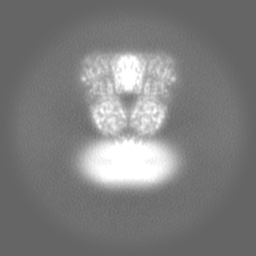
| Projections & slices | Image control
Images are generated by Spider. | ||||||||||||||||||||||||||||||||||||
| Voxel size | X=Y=Z: 1.33 Å | ||||||||||||||||||||||||||||||||||||
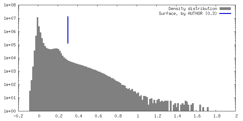
| Density |
| ||||||||||||||||||||||||||||||||||||
| Symmetry | Space group: 1 | ||||||||||||||||||||||||||||||||||||
| Details | EMDB XML:
|
 Z (Sec.)
 Y (Row.)
 X (Col.)




-Supplemental data
- Sample components
-Entire : Heteromeric GluA1A2 with competitive antagonist ZK
| Entire | Name: Heteromeric GluA1A2 with competitive antagonist ZK |
|---|---|
| Components |
|
-Supramolecule #1: Heteromeric GluA1A2 with competitive antagonist ZK
| Supramolecule | Name: Heteromeric GluA1A2 with competitive antagonist ZK / type: complex / ID: 1 / Parent: 0 / Macromolecule list: #1-#2 |
|---|---|
| Source (natural) | Organism: Rattus norvegicus (Norway rat) |
| Molecular weight | Theoretical: 556 KDa |
-Macromolecule #1: Isoform Flip of Glutamate receptor 1
| Macromolecule | Name: Isoform Flip of Glutamate receptor 1 / type: protein_or_peptide / ID: 1 / Number of copies: 2 / Enantiomer: LEVO |
|---|---|
| Source (natural) | Organism: Rattus norvegicus (Norway rat) |
| Molecular weight | Theoretical: 91.225055 KDa |
| Recombinant expression | Organism:  Homo sapiens (human) Homo sapiens (human) |
| Sequence | String: NNIQIGGLFP NQQSQEHAAF RFALSQLTEP PKLLPQIDIV NISDSFEMTY RFCSQFSKGV YAIFGFYERR TVNMLTSFCG ALHVCFITP SFPVDTSNQF VLQLRPELQE ALISIIDHYK WQTFVYIYDA DRGLSVLQRV LDTAAEKNWQ VTAVNILTTT E EGYRMLFQ ...String: NNIQIGGLFP NQQSQEHAAF RFALSQLTEP PKLLPQIDIV NISDSFEMTY RFCSQFSKGV YAIFGFYERR TVNMLTSFCG ALHVCFITP SFPVDTSNQF VLQLRPELQE ALISIIDHYK WQTFVYIYDA DRGLSVLQRV LDTAAEKNWQ VTAVNILTTT E EGYRMLFQ DLEKKKERLV VVDCESERLN AILGQIVKLE KNGIGYHYIL ANLGFMDIDL NKFKESGANV TGFQLVNYTD TI PARIMQQ WRTSDSRDHT RVDWKRPKYT SALTYDGVKV MAEAFQSLRR QRIDISRRGN AGDCLANPAV PWGQGIDIQR ALQ QVRFEG LTGNVQFNEK GRRTNYTLHV IEMKHDGIRK IGYWNEDDKF VPAGGDNSSV QNRTYIVTTI LEDPYVMLKK NANQ FEGND RYEGYCVELA AEIAKHVGYS YRLEIVSDGK YGARDPDTKA WNGMVGELVY GRADVAVAPL TITLVREEVI DFSKP FMSL GISIMIKKPQ KSKPGVFSFL DPLAYEIWMC IVFAYIGVSV VLFLVSRFSP YEWHSEEFEE GRDQTTSDQS NEFGIF NSL WFSLGAFMQQ GCDISPRSLS GRIVGGVWWF FTLIIISSYT ANLAAFLTVE RMVSPIESAE DLAKQTEIAY GTLEAGS TK EFFRRSKIAV FEKMWTYMKS AEPSVFVRTT EEGMIRVRKS KGKYAYLLES TMNEYIEQRK PCDTMKVGGN LDSKGYGI A TPKGSALRGP VNLAVLKLSE QGVLDKLKSK WWYDKGECGS KDSGSKDKTS ALSLSNVAGV FYILIGGLGL AMLVALIEF CYKSR UniProtKB: Glutamate receptor 1 |
-Macromolecule #2: Isoform Flip of Glutamate receptor 2
| Macromolecule | Name: Isoform Flip of Glutamate receptor 2 / type: protein_or_peptide / ID: 2 / Number of copies: 2 / Enantiomer: LEVO |
|---|---|
| Source (natural) | Organism: Rattus norvegicus (Norway rat) |
| Molecular weight | Theoretical: 91.054867 KDa |
| Recombinant expression | Organism: Homo sapiens (human) |
| Sequence | String: NSIQIGGLFP RGADQEYSAF RVGMVQFSTS EFRLTPHIDN LEVANSFAVT NAFCSQFSRG VYAIFGFYDK KSVNTITSFC GTLHVSFIT PSFPTDGTHP FVIQMRPDLK GALLSLIEYY QWDKFAYLYD SDRGLSTLQA VLDSAAEKKW QVTAINVGNI N NDKKDETY ...String: NSIQIGGLFP RGADQEYSAF RVGMVQFSTS EFRLTPHIDN LEVANSFAVT NAFCSQFSRG VYAIFGFYDK KSVNTITSFC GTLHVSFIT PSFPTDGTHP FVIQMRPDLK GALLSLIEYY QWDKFAYLYD SDRGLSTLQA VLDSAAEKKW QVTAINVGNI N NDKKDETY RSLFQDLELK KERRVILDCE RDKVNDIVDQ VITIGKHVKG YHYIIANLGF TDGDLLKIQF GGAEVSGFQI VD YDDSLVS KFIERWSTLE EKEYPGAHTA TIKYTSALTY DAVQVMTEAF RNLRKQRIEI SRRGNAGDCL ANPAVPWGQG VEI ERALKQ VQVEGLSGNI KFDQNGKRIN YTINIMELKT NGPRKIGYWS EVDKMVLTED DTSGLEQKTV VVTTILESPY VMMK KNHEM LEGNERYEGY CVDLAAEIAK HCGFKYKLTI VGDGKYGARD ADTKIWNGMV GELVYGKADI AIAPLTITLV REEVI DFSK PFMSLGISIM IKKPQKSKPG VFSFLDPLAY EIWMCIVFAY IGVSVVLFLV SRFSPYEWHT EEFEDGRETW SSESTN EFG IFNSLWFSLG AFMRQGCDIS PRSLSGRIVG GVWWFFTLII ISSYTANLAA FLTVERMVSP IESAEDLSKQ TEIAYGT LD SGSTKEFFRR SKIAVFDKMW TYMRSAEPSV FVRTTAEGVA RVRKSKGKYA YLLESTMNEY IEQRKPCDTM KVGGNLDS K GYGIATPKGS SLGTPVNLAV LKLSEQGVLD KLKNKWWYDK GECGAKDSGS KEKTSALSLS NVAGVFYILV GGLGLAMLV ALIEFCYKSR A UniProtKB: Glutamate receptor 2 |
-Macromolecule #3: (S)-2-AMINO-3-(3,5-DIOXO-[1,2,4]OXADIAZOLIDIN-2-YL)-PROPIONIC ACID
| Macromolecule | Name: (S)-2-AMINO-3-(3,5-DIOXO-[1,2,4]OXADIAZOLIDIN-2-YL)-PROPIONIC ACID type: ligand / ID: 3 / Number of copies: 4 / Formula: QUS |
|---|---|
| Molecular weight | Theoretical: 189.126 Da |
| Chemical component information | 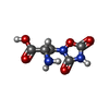 ChemComp-QUS: |
-Experimental details
-Structure determination
| Method | cryo EM |
|---|---|
 Processing Processing | single particle reconstruction |
| Aggregation state | particle |
-Sample preparation
| Concentration | 4.5 mg/mL | |||||||||||||||
|---|---|---|---|---|---|---|---|---|---|---|---|---|---|---|---|---|
| Buffer | pH: 8 Component:
Details: 150 mM NaCl, 20 mM Tris-HCl pH 8.0, and 0.05% digitonin, 1 mM quiqualate | |||||||||||||||
| Grid | Model: UltrAuFoil R1.2/1.3 / Material: GOLD / Mesh: 300 / Support film - Material: GOLD / Support film - topology: HOLEY / Support film - Film thickness: 500 | |||||||||||||||
| Vitrification | Cryogen name: ETHANE / Chamber humidity: 100 % / Chamber temperature: 298 K / Instrument: FEI VITROBOT MARK IV | |||||||||||||||
| Details | The sample had compositional heterogeneity, with broken particles seen throughout. Otherwise, sample was monodisperse |
- Electron microscopy
| Microscope | TFS KRIOS |
|---|---|
| Image recording | Film or detector model: GATAN K3 BIOQUANTUM (6k x 4k) / Average electron dose: 45.7 e/Å2 |
| Electron beam | Acceleration voltage: 300 kV / Electron source:  FIELD EMISSION GUN FIELD EMISSION GUN |
| Electron optics | C2 aperture diameter: 100.0 µm / Illumination mode: SPOT SCAN / Imaging mode: BRIGHT FIELD / Cs: 2.7 mm / Nominal defocus max: 2.5 µm / Nominal defocus min: 1.0 µm |
| Experimental equipment |  Model: Titan Krios / Image courtesy: FEI Company |
