 Movie
Movie Controller


+ Open data

- Basic information
| Entry | Database: EMDB / ID: EMD-6713 | |||||||||
|---|---|---|---|---|---|---|---|---|---|---|
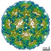
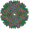
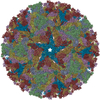
| Title | Inner capsid of Rice Dwarf Virus with T-trimers | |||||||||
 Map data Map data | ||||||||||
 Sample Sample |
| |||||||||
| Biological species |  Rice dwarf virus (isolate O) Rice dwarf virus (isolate O) | |||||||||
| Method | single particle reconstruction / cryo EM / Resolution: 13.43 Å | |||||||||
 Authors Authors | Nakamichi Y / Miyazaki N / Tsutsumi K / Higashiura A / Murata K / Nakagawa A | |||||||||
 Citation Citation | Journal: Structure / Year: 2019 Title: An Assembly Intermediate Structure of Rice Dwarf Virus Reveals a Hierarchical Outer Capsid Shell Assembly Mechanism. Authors: Yusuke Nakamichi / Naoyuki Miyazaki / Kenta Tsutsumi / Akifumi Higashiura / Hirotaka Narita / Kazuyoshi Murata / Atsushi Nakagawa /  Abstract: Nearly all viruses of the Reoviridae family possess a multi-layered capsid consisting of an inner layer with icosahedral T = 1 symmetry and a second-outer layer (composed of 260 copies of a trimeric ...Nearly all viruses of the Reoviridae family possess a multi-layered capsid consisting of an inner layer with icosahedral T = 1 symmetry and a second-outer layer (composed of 260 copies of a trimeric protein) exhibiting icosahedral T = 13 symmetry. Here we describe the construction and structural evaluation of an assembly intermediate of the Rice dwarf virus of the family Reoviridae stalled at the second capsid layer via targeted disruption of the trimer-trimer interaction interface in the second-layer capsid protein. Structural determination was performed by conventional and Zernike/Volta phase-contrast cryoelectron microscopy. The assembly defect second-layer capsid trimers bound exclusively to the outer surface of the innermost capsid layer at the icosahedral 3-fold axis. Furthermore, the second-layer assembly could not proceed without specific inter-trimer interactions. Our results suggest that the correct assembly pathway for second-layer capsid formation is highly controlled at the inter-layer and inter-trimer interactions. | |||||||||
| History |
|
- Structure visualization
| Movie |
 Movie viewer Movie viewer |
|---|---|
| Structure viewer | EM map: SurfViewMolmilJmol/JSmol |
| Supplemental images |

- Downloads & links
-EMDB archive
| Map data | emd_6713.map.gz | 216.6 MB | EMDB map data format | |
|---|---|---|---|---|
| Header (meta data) | emd-6713-v30.xmlemd-6713.xml | 11.5 KB 11.5 KB | Display Display | EMDB header |
| Images | 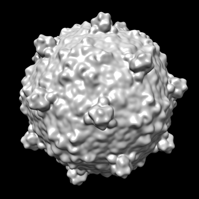 emd_6713.png emd_6713.png | 71.6 KB | ||
| Archive directory |  http://ftp.pdbj.org/pub/emdb/structures/EMD-6713ftp://ftp.pdbj.org/pub/emdb/structures/EMD-6713 http://ftp.pdbj.org/pub/emdb/structures/EMD-6713ftp://ftp.pdbj.org/pub/emdb/structures/EMD-6713 | HTTPS FTP |
-Related structure data
-Links
| EMDB pages | EMDB (EBI/PDBe) / EMDataResource |
|---|
-Map
| File | Download / File: emd_6713.map.gz / Format: CCP4 / Size: 244.1 MB / Type: IMAGE STORED AS FLOATING POINT NUMBER (4 BYTES) | ||||||||||||||||||||||||||||||||||||||||||||||||||||||||||||
|---|---|---|---|---|---|---|---|---|---|---|---|---|---|---|---|---|---|---|---|---|---|---|---|---|---|---|---|---|---|---|---|---|---|---|---|---|---|---|---|---|---|---|---|---|---|---|---|---|---|---|---|---|---|---|---|---|---|---|---|---|---|
| Projections & slices | Image control
Images are generated by Spider. | ||||||||||||||||||||||||||||||||||||||||||||||||||||||||||||
| Voxel size | X=Y=Z: 1.88 Å | ||||||||||||||||||||||||||||||||||||||||||||||||||||||||||||
| Density |
| ||||||||||||||||||||||||||||||||||||||||||||||||||||||||||||
| Symmetry | Space group: 1 | ||||||||||||||||||||||||||||||||||||||||||||||||||||||||||||
| Details | EMDB XML:
CCP4 map header:
| ||||||||||||||||||||||||||||||||||||||||||||||||||||||||||||
 Z (Sec.)
 Y (Row.)
 X (Col.)


-Supplemental data
- Sample components
-Entire : Rice dwarf virus (isolate O)
| Entire | Name: Rice dwarf virus (isolate O) |
|---|---|
| Components |
|
-Supramolecule #1: Rice dwarf virus (isolate O)
| Supramolecule | Name: Rice dwarf virus (isolate O) / type: virus / ID: 1 / Parent: 0 / Macromolecule list: #1 / NCBI-ID: 142805 / Sci species name: Rice dwarf virus (isolate O) / Virus type: VIRION / Virus isolate: SPECIES / Virus enveloped: No / Virus empty: No |
|---|---|
| Host (natural) | Organism:  |
| Virus shell | Shell ID: 1 / Name: inner shell / T number (triangulation number): 1 |
| Virus shell | Shell ID: 2 / Name: outer shell / T number (triangulation number): 13 |
-Experimental details
-Structure determination
| Method | cryo EM |
|---|---|
 Processing Processing | single particle reconstruction |
| Aggregation state | particle |
-Sample preparation
| Buffer | pH: 7.4 Component:
| |||||||||||||||
|---|---|---|---|---|---|---|---|---|---|---|---|---|---|---|---|---|
| Vitrification | Cryogen name: ETHANE |
- Electron microscopy
| Microscope | JEOL 2200FS |
|---|---|
| Specialist optics | Energy filter - Name: omega / Energy filter - Lower energy threshold: 0 eV / Energy filter - Upper energy threshold: 20 eV |
| Image recording | Film or detector model: TVIPS TEMCAM-F415 (4k x 4k) / Digitization - Dimensions - Width: 4096 pixel / Digitization - Dimensions - Height: 4096 pixel / Average exposure time: 1.0 sec. / Average electron dose: 20.0 e/Å2 |
| Electron beam | Acceleration voltage: 200 kV / Electron source:  FIELD EMISSION GUN FIELD EMISSION GUN |
| Electron optics | Illumination mode: FLOOD BEAM / Imaging mode: BRIGHT FIELD |
| Sample stage | Specimen holder model: GATAN 626 SINGLE TILT LIQUID NITROGEN CRYO TRANSFER HOLDER Cooling holder cryogen: NITROGEN |
