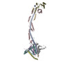
Complex: Bovine F-ATP synthase in the oligomeric form of an IF1 bound planar dimer
Protein or peptide: x 16 types
Keywords
ATP synthase/hydrolase / oligomer / membrane bending / mammalian mitochondria / MEMBRANE PROTEIN
Function / homology
Function and homology information
negative regulation of mitochondrial ATP synthesis coupled proton transport / angiostatin binding / Formation of ATP by chemiosmotic coupling / Cristae formation / ATPase inhibitor activity / mitochondrial proton-transporting ATP synthase complex assembly / mitochondrial envelope / Mitochondrial translation termination / proton channel activity / negative regulation of hydrolase activity ...negative regulation of mitochondrial ATP synthesis coupled proton transport / angiostatin binding / Formation of ATP by chemiosmotic coupling / Cristae formation / ATPase inhibitor activity / mitochondrial proton-transporting ATP synthase complex assembly / mitochondrial envelope / Mitochondrial translation termination / proton channel activity / negative regulation of hydrolase activity / Mitochondrial protein degradation / negative regulation of endothelial cell proliferation / heme biosynthetic process / proton transmembrane transporter activity / proton motive force-driven ATP synthesis / proton-transporting two-sector ATPase complex, proton-transporting domain / proton motive force-driven mitochondrial ATP synthesis / positive regulation of blood vessel endothelial cell migration / MHC class I protein binding / H+-transporting two-sector ATPase / proton-transporting ATP synthase complex / proton-transporting ATP synthase activity, rotational mechanism / proton transmembrane transport / erythrocyte differentiation / aerobic respiration / ADP binding / mitochondrial membrane / ATPase binding / calmodulin binding / protein homotetramerization / mitochondrial inner membrane / structural molecule activity / cell surface / protein homodimerization activity / ATP hydrolysis activity / protein-containing complex / mitochondrion / ATP binding / identical protein binding / plasma membrane Similarity search - Function
ATP synthase, F0 complex, subunit G, mitochondrial / ATP synthase, F0 complex, subunit E, mitochondrial / Mitochondrial ATP synthase subunit g, animal / Mitochondrial ATP synthase g subunit / ATP synthase E chain / ATP synthase protein 8, metazoa / Mitochondrial F1-F0 ATP synthase subunit F, predicted / ATP synthase protein 8, mammals / ATP synthase protein 8 / Mitochondrial F1F0-ATP synthase, subunit f ...ATP synthase, F0 complex, subunit G, mitochondrial / ATP synthase, F0 complex, subunit E, mitochondrial / Mitochondrial ATP synthase subunit g, animal / Mitochondrial ATP synthase g subunit / ATP synthase E chain / ATP synthase protein 8, metazoa / Mitochondrial F1-F0 ATP synthase subunit F, predicted / ATP synthase protein 8, mammals / ATP synthase protein 8 / Mitochondrial F1F0-ATP synthase, subunit f / Mitochondrial ATPase inhibitor / Mitochondrial ATPase inhibitor, IATP / ATP synthase-coupling factor 6, mitochondrial / ATP synthase-coupling factor 6 superfamily, mitochondrial / Mitochondrial ATP synthase coupling factor 6 / : / Metazoan delta subunit of F1F0-ATP synthase, C-terminal domain / ATP synthase, F0 complex, subunit B/MI25 / ATP synthase, F0 complex, subunit B / Mitochondrial ATP synthase B chain precursor (ATP-synt_B) / ATP synthase, F0 complex, subunit D, mitochondrial / ATP synthase D chain, mitochondrial (ATP5H) / ATP synthase delta/epsilon subunit, C-terminal domain superfamily / ATP synthase, F0 complex, subunit D superfamily, mitochondrial / ATP synthase, F0 complex, subunit A, bacterial/mitochondria / ATP synthase, F1 complex, epsilon subunit, mitochondrial / ATP synthase, F1 complex, epsilon subunit superfamily, mitochondrial / Mitochondrial ATP synthase epsilon chain / ATPase, OSCP/delta subunit, conserved site / ATP synthase delta (OSCP) subunit signature. / F1F0 ATP synthase OSCP/delta subunit, N-terminal domain superfamily / ATP synthase, F0 complex, subunit A / ATP synthase, F0 complex, subunit A, active site / ATP synthase, F0 complex, subunit A superfamily / ATP synthase A chain / ATP synthase a subunit signature. / ATPase, OSCP/delta subunit / ATP synthase delta (OSCP) subunit / ATP synthase, F1 complex, delta/epsilon subunit / ATP synthase, F1 complex, delta/epsilon subunit, N-terminal / F0F1 ATP synthase delta/epsilon subunit, N-terminal / ATP synthase, Delta/Epsilon chain, beta-sandwich domain / ATP synthase, F0 complex, subunit C / F1F0 ATP synthase subunit C superfamily / ATP synthase, F0 complex, subunit C, DCCD-binding site / ATP synthase c subunit signature. / ATP synthase, F1 complex, gamma subunit conserved site / ATP synthase gamma subunit signature. / ATP synthase, F1 complex, beta subunit / ATP synthase, alpha subunit, C-terminal domain superfamily / ATP synthase, F1 complex, alpha subunit nucleotide-binding domain / ATP synthase, F1 complex, gamma subunit / ATP synthase, F1 complex, gamma subunit superfamily / ATP synthase / ATP synthase, alpha subunit, C-terminal / ATP synthase, F1 complex, alpha subunit / : / ATP synthase alpha/beta chain, C terminal domain / V-ATPase proteolipid subunit C-like domain / F/V-ATP synthase subunit C superfamily / ATP synthase subunit C / : / ATPase, F1/V1 complex, beta/alpha subunit, C-terminal / C-terminal domain of V and A type ATP synthase / ATPase, F1/V1/A1 complex, alpha/beta subunit, N-terminal domain superfamily / ATP synthase subunit alpha, N-terminal domain-like superfamily / ATPase, F1/V1/A1 complex, alpha/beta subunit, N-terminal domain / ATP synthase alpha/beta family, beta-barrel domain / ATPase, alpha/beta subunit, nucleotide-binding domain, active site / ATP synthase alpha and beta subunits signature. / ATPase, F1/V1/A1 complex, alpha/beta subunit, nucleotide-binding domain / ATP synthase alpha/beta family, nucleotide-binding domain / ATPases associated with a variety of cellular activities / AAA+ ATPase domain / P-loop containing nucleoside triphosphate hydrolase Similarity search - Domain/homology
ATP synthase subunit alpha / ATP synthase F(1) complex catalytic subunit beta, mitochondrial / ATP synthase F(0) complex subunit a / ATPase inhibitor, mitochondrial / ATP synthase peripheral stalk subunit F6, mitochondrial / ATP synthase F(0) complex subunit 8 / ATP synthase F(1) complex subunit delta, mitochondrial / ATP synthase F(1) complex subunit gamma, mitochondrial / ATP synthase F(1) complex subunit epsilon, mitochondrial / ATP synthase F(0) complex subunit C2, mitochondrial ...ATP synthase subunit alpha / ATP synthase F(1) complex catalytic subunit beta, mitochondrial / ATP synthase F(0) complex subunit a / ATPase inhibitor, mitochondrial / ATP synthase peripheral stalk subunit F6, mitochondrial / ATP synthase F(0) complex subunit 8 / ATP synthase F(1) complex subunit delta, mitochondrial / ATP synthase F(1) complex subunit gamma, mitochondrial / ATP synthase F(1) complex subunit epsilon, mitochondrial / ATP synthase F(0) complex subunit C2, mitochondrial / ATP synthase peripheral stalk subunit b, mitochondrial / ATP synthase peripheral stalk subunit d, mitochondrial / ATP synthase peripheral stalk subunit OSCP, mitochondrial / ATP synthase F(0) complex subunit e, mitochondrial / ATP synthase F(0) complex subunit f, mitochondrial / ATP synthase F(0) complex subunit g, mitochondrial Similarity search - Component
Biological species
Bos taurus (domestic cattle)
Method
single particle reconstruction / cryo EM / Resolution: 5.0 Å
Japan Agency for Medical Research and Development (AMED)
JP22ama 121003
Japan
Japan Society for the Promotion of Science (JSPS)
23H02453
Japan
Citation
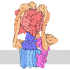
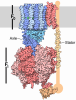
Journal: Cell Death Differ / Year: 2026 Title: A planar dimer of bovine ATP synthase. Authors: Chimari Jiko / Atsuki Nakano / Yosuke Teshirogi / Eiki Yamashita / Genji Kurisu / Daron Standley / Tohru Terada / Kaoru Mitsuoka / Ken Yokoyama / Christoph Gerle / Abstract: Mammalian mitochondrial ATP synthase typically organizes into rows of V-shaped dimers that impose significant membrane curvature essential for mitochondrial cristae formation. Using gentle, column- ...Mammalian mitochondrial ATP synthase typically organizes into rows of V-shaped dimers that impose significant membrane curvature essential for mitochondrial cristae formation. Using gentle, column-free purification combined with single-particle cryo-electron microscopy, we have identified a previously unrecognized planar dimeric form of bovine ATP synthase exhibiting minimal membrane bending. This planar dimer is characterized structurally by anti-parallel arrangement of two ATP synthase complexes linked by a straight conformation of inhibitory factor 1 (IF1), a sharp contrast to the kinked IF1 observed in tetrameric assemblies. Molecular dynamics simulations confirm that transitioning between straight and kinked IF1 conformations occurs without significant energetic barriers. The planar dimer also displays distinct peripheral stalk positioning relative to its adjacent α subunit. These structural divergences suggest a specialized function and a distinct localization for planar ATP synthase dimers, providing structural support for a division of labor within mitochondrial ATP synthase populations.
Entire : Bovine F-ATP synthase in the oligomeric form of an IF1 bound plan...
Entire
Name: Bovine F-ATP synthase in the oligomeric form of an IF1 bound planar dimer
Components
Complex: Bovine F-ATP synthase in the oligomeric form of an IF1 bound planar dimer
Protein or peptide: ATP synthase F(0) complex subunit 8
Protein or peptide: ATP synthase subunit alpha
Protein or peptide: ATP synthase F(1) complex catalytic subunit beta, mitochondrial
Protein or peptide: ATP synthase F(1) complex subunit gamma, mitochondrial
Protein or peptide: ATP synthase F(1) complex subunit delta, mitochondrial
Protein or peptide: ATP synthase F(1) complex subunit epsilon, mitochondrial
Protein or peptide: ATPase inhibitor, mitochondrial
Protein or peptide: ATP synthase F(0) complex subunit C2, mitochondrial
Protein or peptide: ATP synthase peripheral stalk subunit OSCP, mitochondrial
Protein or peptide: ATP synthase F(0) complex subunit a
Protein or peptide: ATP synthase peripheral stalk subunit b, mitochondrial
Protein or peptide: ATP synthase peripheral stalk subunit d, mitochondrial
Protein or peptide: ATP synthase F(0) complex subunit e, mitochondrial
Protein or peptide: ATP synthase F(0) complex subunit f, mitochondrial
Protein or peptide: ATP synthase F(0) complex subunit g, mitochondrial
Protein or peptide: ATP synthase peripheral stalk subunit F6, mitochondrial
+
Supramolecule #1: Bovine F-ATP synthase in the oligomeric form of an IF1 bound plan...
Supramolecule
Name: Bovine F-ATP synthase in the oligomeric form of an IF1 bound planar dimer type: complex / ID: 1 / Parent: 0 / Macromolecule list: all Details: Oligomer fractions of bovine F-ATP synthase obtained from heart muscle tissue mitochondria.
UniProtKB: ATP synthase peripheral stalk subunit F6, mitochondrial
-
Experimental details
-
Structure determination
Method
cryo EM
Processing
single particle reconstruction
Aggregation state
particle
-
Sample preparation
Concentration
5 mg/mL
Buffer
pH: 7.3
Grid
Model: Quantifoil R2/2 / Material: COPPER / Mesh: 300 / Support film - Material: CARBON / Support film - topology: CONTINUOUS / Support film - Film thickness: 2 / Pretreatment - Type: GLOW DISCHARGE / Pretreatment - Time: 30 sec.
Vitrification
Cryogen name: ETHANE / Chamber humidity: 100 % / Chamber temperature: 277 K / Instrument: FEI VITROBOT MARK IV
-
Electron microscopy
Microscope
TFS KRIOS
Image recording
Film or detector model: GATAN K3 (6k x 4k) / Average electron dose: 50.0 e/Å2
Electron beam
Acceleration voltage: 300 kV / Electron source: FIELD EMISSION GUN
In the structure databanks used in Yorodumi, some data are registered as the other names, "COVID-19 virus" and "2019-nCoV". Here are the details of the virus and the list of structure data.
Jan 31, 2019. EMDB accession codes are about to change! (news from PDBe EMDB page)
EMDB accession codes are about to change! (news from PDBe EMDB page)
The allocation of 4 digits for EMDB accession codes will soon come to an end. Whilst these codes will remain in use, new EMDB accession codes will include an additional digit and will expand incrementally as the available range of codes is exhausted. The current 4-digit format prefixed with “EMD-” (i.e. EMD-XXXX) will advance to a 5-digit format (i.e. EMD-XXXXX), and so on. It is currently estimated that the 4-digit codes will be depleted around Spring 2019, at which point the 5-digit format will come into force.
The EM Navigator/Yorodumi systems omit the EMD- prefix.
Related info.:Q: What is EMD? / ID/Accession-code notation in Yorodumi/EM Navigator
Yorodumi is a browser for structure data from EMDB, PDB, SASBDB, etc.
This page is also the successor to EM Navigator detail page, and also detail information page/front-end page for Omokage search.
The word "yorodu" (or yorozu) is an old Japanese word meaning "ten thousand". "mi" (miru) is to see.
Related info.:EMDB / PDB / SASBDB / Comparison of 3 databanks / Yorodumi Search / Aug 31, 2016. New EM Navigator & Yorodumi / Yorodumi Papers / Jmol/JSmol / Function and homology information / Changes in new EM Navigator and Yorodumi
 Movie
Movie Controller


 Open data
 Basic information
 Map data
 Sample
 Keywords
 Function and homology information
 Bos taurus (domestic cattle)
 Authors
 Japan, 2 items
  Citation
 Structure visualization
 Downloads & links
 emd_65237.png
 https://data.pdbj.org/pub/emdb/structures/EMD-65237


 Z (Sec.)
 Y (Row.)
 X (Col.)


 Sample components
 Processing
 Electron microscopy
 FIELD EMISSION GUN