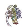
Movie
Movie Controller

[English] 日本語
 Yorodumi
- PDB-9vpd: Cryo-EM structure of the IF1 bound bovine ATP synthase monomer: r... -
+ Open data

- Basic information
| Entry | Database: PDB / ID: 9vpd | |||||||||||||||||||||||||||
|---|---|---|---|---|---|---|---|---|---|---|---|---|---|---|---|---|---|---|---|---|---|---|---|---|---|---|---|---|
| Title | Cryo-EM structure of the IF1 bound bovine ATP synthase monomer: rotary state 1, F1 focused map | |||||||||||||||||||||||||||
 Components Components |
| |||||||||||||||||||||||||||
 Keywords Keywords | MEMBRANE PROTEIN / ATP synthase/hydrolase / oligomer / membrane bending / mammalian mitochondria / motor protein | |||||||||||||||||||||||||||
| Function / homology |  Function and homology information Function and homology informationnegative regulation of mitochondrial ATP synthesis coupled proton transport / angiostatin binding / Formation of ATP by chemiosmotic coupling / Cristae formation / ATPase inhibitor activity / mitochondrial proton-transporting ATP synthase complex assembly / mitochondrial envelope / negative regulation of hydrolase activity / Mitochondrial protein degradation / negative regulation of endothelial cell proliferation ...negative regulation of mitochondrial ATP synthesis coupled proton transport / angiostatin binding / Formation of ATP by chemiosmotic coupling / Cristae formation / ATPase inhibitor activity / mitochondrial proton-transporting ATP synthase complex assembly / mitochondrial envelope / negative regulation of hydrolase activity / Mitochondrial protein degradation / negative regulation of endothelial cell proliferation / heme biosynthetic process / proton transmembrane transporter activity / proton motive force-driven ATP synthesis / proton motive force-driven mitochondrial ATP synthesis / H+-transporting two-sector ATPase / proton-transporting ATP synthase complex / proton-transporting ATP synthase activity, rotational mechanism / proton transmembrane transport / aerobic respiration / erythrocyte differentiation / ADP binding / ATPase binding / calmodulin binding / protein homotetramerization / mitochondrial inner membrane / structural molecule activity / cell surface / protein homodimerization activity / ATP hydrolysis activity / protein-containing complex / mitochondrion / ATP binding / identical protein binding / plasma membrane Similarity search - Function | |||||||||||||||||||||||||||
| Biological species |   Bos taurus (domestic cattle) Bos taurus (domestic cattle) | |||||||||||||||||||||||||||
| Method | ELECTRON MICROSCOPY / single particle reconstruction / cryo EM / Resolution: 2.3 Å | |||||||||||||||||||||||||||
 Authors Authors | Nakano, A. / Jiko, C. / Yamashita, E. / Yokoyama, K. / Gerle, C. | |||||||||||||||||||||||||||
| Funding support |  Japan, 2items Japan, 2items
| |||||||||||||||||||||||||||
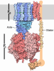
 Citation Citation | Journal: Cell Death Differ / Year: 2026 Title: A planar dimer of bovine ATP synthase. Authors: Chimari Jiko / Atsuki Nakano / Yosuke Teshirogi / Eiki Yamashita / Genji Kurisu / Daron Standley / Tohru Terada / Kaoru Mitsuoka / Ken Yokoyama / Christoph Gerle / Abstract: Mammalian mitochondrial ATP synthase typically organizes into rows of V-shaped dimers that impose significant membrane curvature essential for mitochondrial cristae formation. Using gentle, column- ...Mammalian mitochondrial ATP synthase typically organizes into rows of V-shaped dimers that impose significant membrane curvature essential for mitochondrial cristae formation. Using gentle, column-free purification combined with single-particle cryo-electron microscopy, we have identified a previously unrecognized planar dimeric form of bovine ATP synthase exhibiting minimal membrane bending. This planar dimer is characterized structurally by anti-parallel arrangement of two ATP synthase complexes linked by a straight conformation of inhibitory factor 1 (IF1), a sharp contrast to the kinked IF1 observed in tetrameric assemblies. Molecular dynamics simulations confirm that transitioning between straight and kinked IF1 conformations occurs without significant energetic barriers. The planar dimer also displays distinct peripheral stalk positioning relative to its adjacent α subunit. These structural divergences suggest a specialized function and a distinct localization for planar ATP synthase dimers, providing structural support for a division of labor within mitochondrial ATP synthase populations. #1: Journal: Biorxiv / Year: 2025Title: A planar dimer of bovine ATP synthase Authors: Jiko, C. / Nakano, A. / Teshirogi, Y. / Yamashita, E. / Kurisu, G. / Standley, D. / Terada, T. / Mitsuoka, K. / Yokoyama, K. / Gerle, C. | |||||||||||||||||||||||||||
| History |
|
- Structure visualization
| Structure viewer | Molecule: MolmilJmol/JSmol |
|---|
- Downloads & links
-Download
| PDBx/mmCIF format | 9vpd.cif.gz | 643.1 KB | Display | PDBx/mmCIF format |
|---|---|---|---|---|
| PDB format | pdb9vpd.ent.gz | Display | PDB format | |
| PDBx/mmJSON format | 9vpd.json.gz | Tree view | PDBx/mmJSON format | |
| Others |  Other downloads Other downloads |
-Validation report
| Arichive directory | https://data.pdbj.org/pub/pdb/validation_reports/vp/9vpdftp://data.pdbj.org/pub/pdb/validation_reports/vp/9vpd | HTTPS FTP |
|---|
-Related structure data
| Related structure data |  65239MC  9vpbC  9vpcC M: map data used to model this data C: citing same article ( |
|---|---|
| Similar structure data |


-Links
 PDBj
PDBj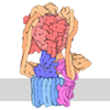
- Assembly
| Deposited unit | 
|
|---|---|
| 1 |
|
-Components
-ATP synthase F(1) complex subunit ... , 4 types, 6 molecules ABCGHI
| #1: Protein | Mass: 59795.492 Da / Num. of mol.: 3 / Source method: isolated from a natural source / Source: (natural) Bos taurus (domestic cattle) / References: UniProt: P19483#3: Protein | | Mass: 33119.035 Da / Num. of mol.: 1 / Source method: isolated from a natural source / Source: (natural) Bos taurus (domestic cattle) / References: UniProt: P05631#4: Protein | | Mass: 17626.992 Da / Num. of mol.: 1 / Source method: isolated from a natural source / Source: (natural) Bos taurus (domestic cattle) / References: UniProt: P05630#5: Protein | | Mass: 5793.889 Da / Num. of mol.: 1 / Source method: isolated from a natural source / Source: (natural) Bos taurus (domestic cattle) / References: UniProt: P05632 |
|---|
-Protein , 2 types, 4 molecules DEFJ
| #2: Protein | Mass: 56340.199 Da / Num. of mol.: 3 / Source method: isolated from a natural source / Source: (natural) Bos taurus (domestic cattle)References: UniProt: P00829, H+-transporting two-sector ATPase #6: Protein | | Mass: 12326.715 Da / Num. of mol.: 1 / Source method: isolated from a natural source / Source: (natural) Bos taurus (domestic cattle) / References: UniProt: P01096 |
|---|
-ATP synthase peripheral stalk subunit ... , 4 types, 4 molecules Sbdh
| #7: Protein | Mass: 23351.596 Da / Num. of mol.: 1 / Source method: isolated from a natural source / Source: (natural) Bos taurus (domestic cattle) / References: UniProt: P13621 |
|---|---|
| #8: Protein | Mass: 28859.629 Da / Num. of mol.: 1 / Source method: isolated from a natural source / Source: (natural) Bos taurus (domestic cattle) / References: UniProt: P13619 |
| #9: Protein | Mass: 18719.453 Da / Num. of mol.: 1 / Source method: isolated from a natural source / Source: (natural) Bos taurus (domestic cattle) / References: UniProt: P13620 |
| #10: Protein | Mass: 12549.321 Da / Num. of mol.: 1 / Source method: isolated from a natural source / Source: (natural) Bos taurus (domestic cattle) / References: UniProt: P02721 |
-Non-polymers , 4 types, 28 molecules 



| #11: Chemical | 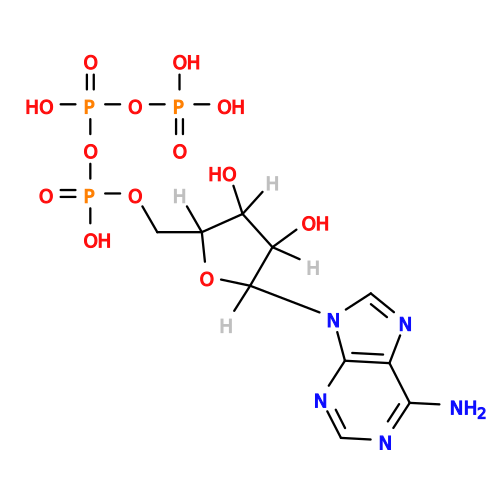 Mass: 507.181 Da / Num. of mol.: 3 / Source method: obtained synthetically / Formula: C10H16N5O13P3 / Feature type: SUBJECT OF INVESTIGATION / Comment: ATP, energy-carrying molecule*YM Mass: 507.181 Da / Num. of mol.: 3 / Source method: obtained synthetically / Formula: C10H16N5O13P3 / Feature type: SUBJECT OF INVESTIGATION / Comment: ATP, energy-carrying molecule*YM#12: Chemical | ChemComp-MG /  Mass: 24.305 Da / Num. of mol.: 5 / Source method: obtained synthetically / Formula: Mg / Feature type: SUBJECT OF INVESTIGATION Mass: 24.305 Da / Num. of mol.: 5 / Source method: obtained synthetically / Formula: Mg / Feature type: SUBJECT OF INVESTIGATION#13: Chemical | 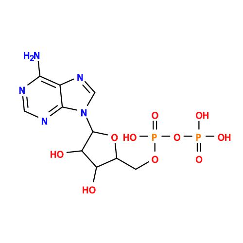 Mass: 427.201 Da / Num. of mol.: 3 / Source method: obtained synthetically / Formula: C10H15N5O10P2 / Feature type: SUBJECT OF INVESTIGATION / Comment: ADP, energy-carrying molecule*YM Mass: 427.201 Da / Num. of mol.: 3 / Source method: obtained synthetically / Formula: C10H15N5O10P2 / Feature type: SUBJECT OF INVESTIGATION / Comment: ADP, energy-carrying molecule*YM#14: Water | ChemComp-HOH / | Mass: 18.015 Da / Num. of mol.: 17 / Source method: isolated from a natural source / Formula: H2O |
|---|
-Details
| Has ligand of interest | Y |
|---|---|
| Has protein modification | N |
-Experimental details
-Experiment
| Experiment | Method: ELECTRON MICROSCOPY |
|---|---|
| EM experiment | Aggregation state: PARTICLE / 3D reconstruction method: single particle reconstruction |
- Sample preparation
| Component | Name: IF1 bound bovine ATP synthase monomer isolated from heart muscle tissue mitochondria Type: COMPLEX / Entity ID: #1-#10 / Source: NATURAL |
|---|---|
| Molecular weight | Value: 0.6 MDa / Experimental value: NO |
| Source (natural) | Organism: Bos taurus (domestic cattle) |
| Buffer solution | pH: 7.3 |
| Specimen | Conc.: 4 mg/ml / Embedding applied: NO / Shadowing applied: NO / Staining applied: NO / Vitrification applied: YES |
| Specimen support | Grid material: COPPER / Grid mesh size: 300 divisions/in. / Grid type: Quantifoil R2/2 |
| Vitrification | Instrument: FEI VITROBOT MARK IV / Cryogen name: ETHANE |
- Electron microscopy imaging
| Experimental equipment |  Model: Titan Krios / Image courtesy: FEI Company |
|---|---|
| Microscopy | Model: TFS KRIOS |
| Electron gun | Electron source:  FIELD EMISSION GUN / Accelerating voltage: 300 kV / Illumination mode: FLOOD BEAM FIELD EMISSION GUN / Accelerating voltage: 300 kV / Illumination mode: FLOOD BEAM |
| Electron lens | Mode: BRIGHT FIELD / Nominal defocus max: 2000 nm / Nominal defocus min: 800 nm / Cs: 2.7 mm |
| Specimen holder | Cryogen: NITROGEN / Specimen holder model: FEI TITAN KRIOS AUTOGRID HOLDER |
| Image recording | Electron dose: 50 e/Å2 / Film or detector model: GATAN K3 (6k x 4k) |
- Processing
| EM software |
| ||||||||||||||||||||||||
|---|---|---|---|---|---|---|---|---|---|---|---|---|---|---|---|---|---|---|---|---|---|---|---|---|---|
| CTF correction | Type: PHASE FLIPPING AND AMPLITUDE CORRECTION | ||||||||||||||||||||||||
| 3D reconstruction | Resolution: 2.3 Å / Resolution method: FSC 0.143 CUT-OFF / Num. of particles: 398620 / Algorithm: FOURIER SPACE / Symmetry type: POINT | ||||||||||||||||||||||||
| Atomic model building | Protocol: FLEXIBLE FIT / Space: REAL | ||||||||||||||||||||||||
| Atomic model building | PDB-ID: 6YY0 Accession code: 6YY0 / Source name: PDB / Type: experimental model |