 ムービー
ムービー コントローラー


+ データを開く

- 基本情報
基本情報
| 登録情報 | データベース: EMDB / ID: EMD-6211 | |||||||||
|---|---|---|---|---|---|---|---|---|---|---|
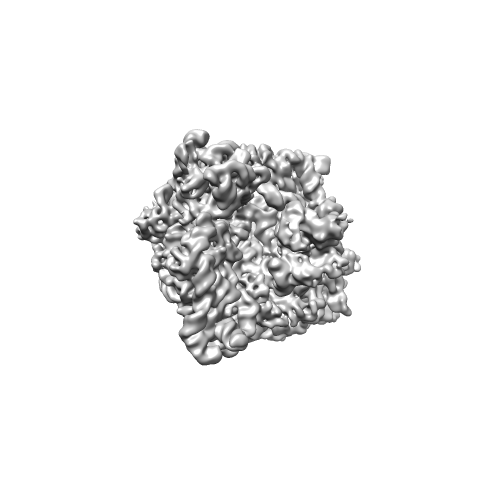
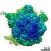
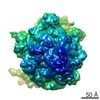
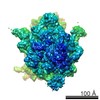
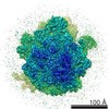
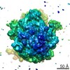
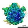
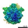
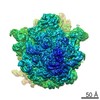
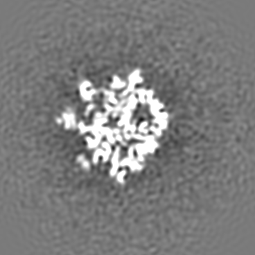
| タイトル | Cryo-EM structure of ribosomal protein S1 on the ribosome | |||||||||
 マップデータ マップデータ | Reconstruction of ErmCL-stalled E. coli ribosome containing density for the first domains of ribosomal protein S1 | |||||||||
 試料 試料 |
| |||||||||
 キーワード キーワード | Ribosome / Ribosomal protein S1 / Cryo-EM | |||||||||
| 生物種 |  | |||||||||
| 手法 | 単粒子再構成法 / クライオ電子顕微鏡法 / 解像度: 9.5 Å | |||||||||
 データ登録者 データ登録者 | Byrgazov K / Grishkovskaya I / Arenz S / Coudevylle N / Temmel H / Wilson DN / Djinovic-Carugo K / Moll I | |||||||||
 引用 引用 | ジャーナル: Nucleic Acids Res / 年: 2015 タイトル: Structural basis for the interaction of protein S1 with the Escherichia coli ribosome. 著者: Konstantin Byrgazov / Irina Grishkovskaya / Stefan Arenz / Nicolas Coudevylle / Hannes Temmel / Daniel N Wilson / Kristina Djinovic-Carugo / Isabella Moll /    要旨: In Gram-negative bacteria, the multi-domain protein S1 is essential for translation initiation, as it recruits the mRNA and facilitates its localization in the decoding centre. In sharp contrast to ...In Gram-negative bacteria, the multi-domain protein S1 is essential for translation initiation, as it recruits the mRNA and facilitates its localization in the decoding centre. In sharp contrast to its functional importance, S1 is still lacking from the high-resolution structures available for Escherichia coli and Thermus thermophilus ribosomes and thus the molecular mechanism governing the S1-ribosome interaction has still remained elusive. Here, we present the structure of the N-terminal S1 domain D1 when bound to the ribosome at atomic resolution by using a combination of NMR, X-ray crystallography and cryo-electron microscopy. Together with biochemical assays, the structure reveals that S1 is anchored to the ribosome primarily via a stabilizing π-stacking interaction within the short but conserved N-terminal segment that is flexibly connected to domain D1. This interaction is further stabilized by salt bridges involving the zinc binding pocket of protein S2. Overall, this work provides one hitherto enigmatic piece in the 'ribosome puzzle', namely the detailed molecular insight into the topology of the S1-ribosome interface. Moreover, our data suggest novel mechanisms that have the potential to modulate protein synthesis in response to environmental cues by changing the affinity of S1 for the ribosome. | |||||||||
| 履歴 |
|
- 構造の表示
構造の表示
| ムービー |
 ムービービューア ムービービューア |
|---|---|
| 構造ビューア | EMマップ: SurfViewMolmilJmol/JSmol |
| 添付画像 |
- ダウンロードとリンク
-EMDBアーカイブ
| マップデータ | emd_6211.map.gz | 177.1 MB | EMDBマップデータ形式 | |
|---|---|---|---|---|
| ヘッダ (付随情報) | emd-6211-v30.xmlemd-6211.xml | 8.7 KB 8.7 KB | 表示 表示 | EMDBヘッダ |
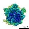
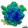
| 画像 |  emd_6211.png emd_6211.png | 61.5 KB | ||
| アーカイブディレクトリ |  http://ftp.pdbj.org/pub/emdb/structures/EMD-6211ftp://ftp.pdbj.org/pub/emdb/structures/EMD-6211 http://ftp.pdbj.org/pub/emdb/structures/EMD-6211ftp://ftp.pdbj.org/pub/emdb/structures/EMD-6211 | HTTPS FTP |
-検証レポート
| 文書・要旨 | emd_6211_validation.pdf.gz | 78.3 KB | 表示 | EMDB検証レポート |
|---|---|---|---|---|
| 文書・詳細版 | emd_6211_full_validation.pdf.gz | 77.4 KB | 表示 | |
| XML形式データ | emd_6211_validation.xml.gz | 493 B | 表示 | |
| アーカイブディレクトリ | https://ftp.pdbj.org/pub/emdb/validation_reports/EMD-6211ftp://ftp.pdbj.org/pub/emdb/validation_reports/EMD-6211 | HTTPS FTP |
-関連構造データ
-リンク
| EMDBのページ | EMDB (EBI/PDBe) / EMDataResource |
|---|---|
| 「今月の分子」の関連する項目 |

-マップ
| ファイル | ダウンロード / ファイル: emd_6211.map.gz / 形式: CCP4 / 大きさ: 185.7 MB / タイプ: IMAGE STORED AS FLOATING POINT NUMBER (4 BYTES) | ||||||||||||||||||||||||||||||||||||||||||||||||||||||||||||||||||||
|---|---|---|---|---|---|---|---|---|---|---|---|---|---|---|---|---|---|---|---|---|---|---|---|---|---|---|---|---|---|---|---|---|---|---|---|---|---|---|---|---|---|---|---|---|---|---|---|---|---|---|---|---|---|---|---|---|---|---|---|---|---|---|---|---|---|---|---|---|---|
| 注釈 | Reconstruction of ErmCL-stalled E. coli ribosome containing density for the first domains of ribosomal protein S1 | ||||||||||||||||||||||||||||||||||||||||||||||||||||||||||||||||||||
| 投影像・断面図 | 画像のコントロール
画像は Spider により作成 | ||||||||||||||||||||||||||||||||||||||||||||||||||||||||||||||||||||
| ボクセルのサイズ | X=Y=Z: 1.0489 Å | ||||||||||||||||||||||||||||||||||||||||||||||||||||||||||||||||||||
| 密度 |
| ||||||||||||||||||||||||||||||||||||||||||||||||||||||||||||||||||||
| 対称性 | 空間群: 1 | ||||||||||||||||||||||||||||||||||||||||||||||||||||||||||||||||||||
| 詳細 | EMDB XML:
CCP4マップ ヘッダ情報:
| ||||||||||||||||||||||||||||||||||||||||||||||||||||||||||||||||||||
 Z (Sec.)
 Y (Row.)
 X (Col.)


-添付データ
- 試料の構成要素
-全体 : Reconstruction of ErmCL-stalled E. coli ribosome containing densi...
| 全体 | 名称: Reconstruction of ErmCL-stalled E. coli ribosome containing density for the first domains of ribosomal protein S1 |
|---|---|
| 要素 |
|
-超分子 #1000: Reconstruction of ErmCL-stalled E. coli ribosome containing densi...
| 超分子 | 名称: Reconstruction of ErmCL-stalled E. coli ribosome containing density for the first domains of ribosomal protein S1 タイプ: sample / ID: 1000 / Number unique components: 1 |
|---|
-超分子 #1: ErmCL-stalled ribosome
| 超分子 | 名称: ErmCL-stalled ribosome / タイプ: complex / ID: 1 / Name.synonym: 70S ribosome / 組換発現: No / データベース: NCBI / Ribosome-details: ribosome-prokaryote: ALL |
|---|---|
| 由来(天然) | 生物種: |
-実験情報
-構造解析
| 手法 | クライオ電子顕微鏡法 |
|---|---|
 解析 解析 | 単粒子再構成法 |
| 試料の集合状態 | particle |
-試料調製
| 緩衝液 | pH: 7.4 / 詳細: 50 mM HEPES, 100 mM KOAc, 20 mM MgOAc |
|---|---|
| グリッド | 詳細: 2 nm pre-coated Quantifoil R3/3 holey carbon supported grids |
| 凍結 | 凍結剤: ETHANE / 装置: FEI VITROBOT MARK IV |
- 電子顕微鏡法
| 顕微鏡 | FEI TITAN KRIOS |
|---|---|
| 日付 | 2012年5月21日 |
| 撮影 | カテゴリ: CCD フィルム・検出器のモデル: TVIPS TEMCAM-F416 (4k x 4k) 実像数: 6902 / 平均電子線量: 20 e/Å2 |
| 電子線 | 加速電圧: 200 kV / 電子線源:  FIELD EMISSION GUN FIELD EMISSION GUN |
| 電子光学系 | 照射モード: SPOT SCAN / 撮影モード: BRIGHT FIELD / Cs: 2.7 mm / 最大 デフォーカス(公称値): 4.5 µm / 最小 デフォーカス(公称値): 1.2 µm / 倍率(公称値): 75000 |
| 試料ステージ | 試料ホルダーモデル: FEI TITAN KRIOS AUTOGRID HOLDER |
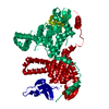
| 実験機器 |  モデル: Titan Krios / 画像提供: FEI Company |
-画像解析
| 詳細 | CTF-parameter estimation in SPIDER TF ED, particle picking in Signature, image processing in SPIDER |
|---|---|
| CTF補正 | 詳細: Defocus groups |
| 最終 再構成 | 解像度のタイプ: BY AUTHOR / 解像度: 9.5 Å / 解像度の算出法: OTHER / ソフトウェア - 名称: SPIDER / 詳細: Final map was filtered to 9.5 Angstrom resolution. / 使用した粒子像数: 128846 |
