 ムービー
ムービー コントローラー


+ データを開く

- 基本情報
基本情報
| 登録情報 | データベース: EMDB / ID: EMD-5947 | |||||||||
|---|---|---|---|---|---|---|---|---|---|---|
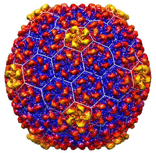
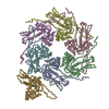
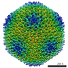
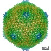
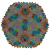
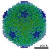
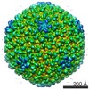
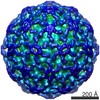
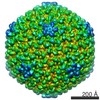
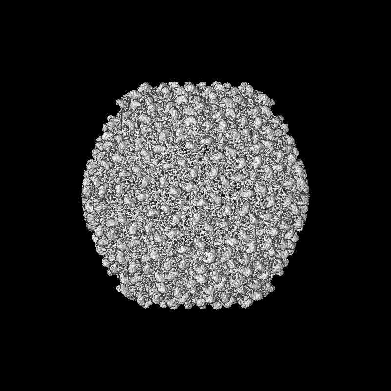
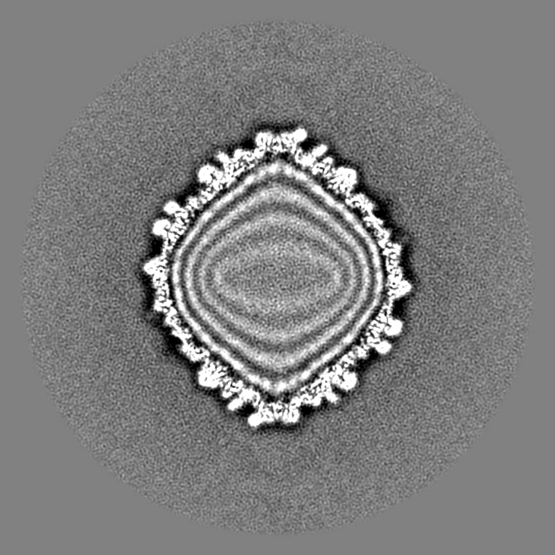
| タイトル | Bacteriophage CUS-3 capsid icosahedral reconstruction | |||||||||
 マップデータ マップデータ | Bacteriophage CUS-3 capsid icosahedral reconstruction | |||||||||
 試料 試料 |
| |||||||||
 キーワード キーワード | mature virion / capsid only / icosahedrally averaged | |||||||||
| 生物種 |  Enterobacteria phage CUS-3 (ファージ) Enterobacteria phage CUS-3 (ファージ) | |||||||||
| 手法 | 単粒子再構成法 / クライオ電子顕微鏡法 / 解像度: 6.8 Å | |||||||||
 データ登録者 データ登録者 | Parent KN / Tang J / Cardone G / Gilcrease EB / Janssen ME / Olson NH / Casjens SR / Baker TS | |||||||||
 引用 引用 | ジャーナル: Virology / 年: 2014 タイトル: Three-dimensional reconstructions of the bacteriophage CUS-3 virion reveal a conserved coat protein I-domain but a distinct tailspike receptor-binding domain. 著者: Kristin N Parent / Jinghua Tang / Giovanni Cardone / Eddie B Gilcrease / Mandy E Janssen / Norman H Olson / Sherwood R Casjens / Timothy S Baker /  要旨: CUS-3 is a short-tailed, dsDNA bacteriophage that infects serotype K1 Escherichia coli. We report icosahedrally averaged and asymmetric, three-dimensional, cryo-electron microscopic reconstructions ...CUS-3 is a short-tailed, dsDNA bacteriophage that infects serotype K1 Escherichia coli. We report icosahedrally averaged and asymmetric, three-dimensional, cryo-electron microscopic reconstructions of the CUS-3 virion. Its coat protein structure adopts the "HK97-fold" shared by other tailed phages and is quite similar to that in phages P22 and Sf6 despite only weak amino acid sequence similarity. In addition, these coat proteins share a unique extra external domain ("I-domain"), suggesting that the group of P22-like phages has evolved over a very long time period without acquiring a new coat protein gene from another phage group. On the other hand, the morphology of the CUS-3 tailspike differs significantly from that of P22 or Sf6, but is similar to the tailspike of phage K1F, a member of the extremely distantly related T7 group of phages. We conclude that CUS-3 obtained its tailspike gene from a distantly related phage quite recently. | |||||||||
| 履歴 |
|
- 構造の表示
構造の表示
| ムービー |
 ムービービューア ムービービューア |
|---|---|
| 構造ビューア | EMマップ: SurfViewMolmilJmol/JSmol |
| 添付画像 |
- ダウンロードとリンク
-EMDBアーカイブ
| マップデータ | emd_5947.map.gz | 602.6 MB | EMDBマップデータ形式 | |
|---|---|---|---|---|
| ヘッダ (付随情報) | emd-5947-v30.xmlemd-5947.xml | 10.3 KB 10.3 KB | 表示 表示 | EMDBヘッダ |
| 画像 |  emd_5947.png emd_5947.png | 147.5 KB | ||
| アーカイブディレクトリ |  http://ftp.pdbj.org/pub/emdb/structures/EMD-5947ftp://ftp.pdbj.org/pub/emdb/structures/EMD-5947 http://ftp.pdbj.org/pub/emdb/structures/EMD-5947ftp://ftp.pdbj.org/pub/emdb/structures/EMD-5947 | HTTPS FTP |
-関連構造データ
-リンク
| EMDBのページ | EMDB (EBI/PDBe) / EMDataResource |
|---|
-マップ
| ファイル | ダウンロード / ファイル: emd_5947.map.gz / 形式: CCP4 / 大きさ: 1.9 GB / タイプ: IMAGE STORED AS FLOATING POINT NUMBER (4 BYTES) | ||||||||||||||||||||||||||||||||||||||||||||||||||||||||||||||||||||
|---|---|---|---|---|---|---|---|---|---|---|---|---|---|---|---|---|---|---|---|---|---|---|---|---|---|---|---|---|---|---|---|---|---|---|---|---|---|---|---|---|---|---|---|---|---|---|---|---|---|---|---|---|---|---|---|---|---|---|---|---|---|---|---|---|---|---|---|---|---|
| 注釈 | Bacteriophage CUS-3 capsid icosahedral reconstruction | ||||||||||||||||||||||||||||||||||||||||||||||||||||||||||||||||||||
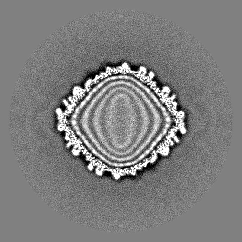
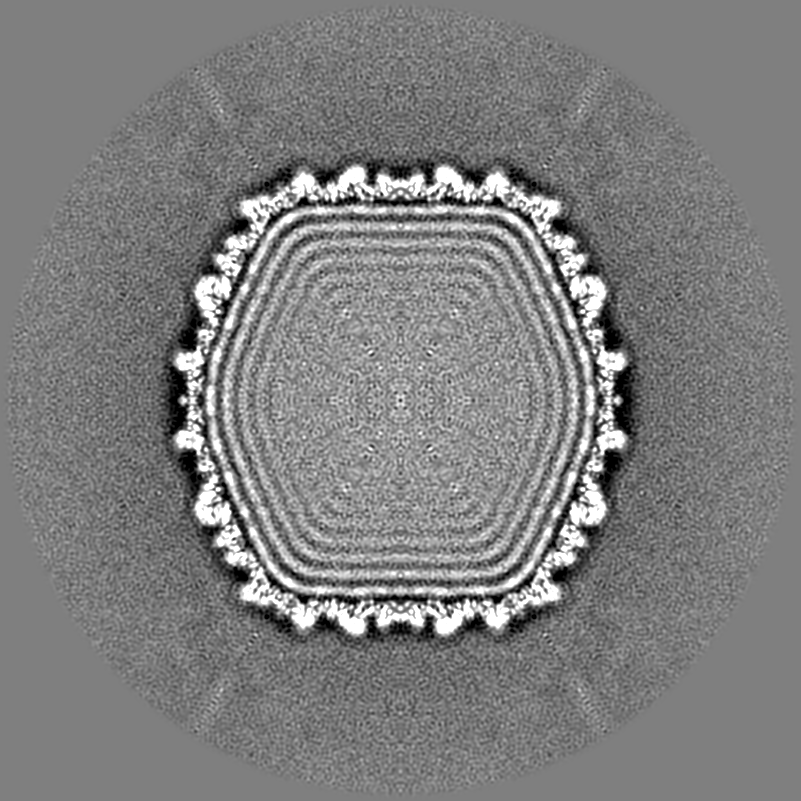
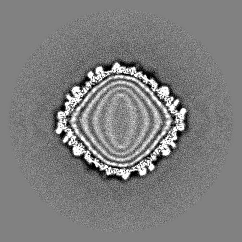
| 投影像・断面図 | 画像のコントロール
画像は Spider により作成 | ||||||||||||||||||||||||||||||||||||||||||||||||||||||||||||||||||||
| ボクセルのサイズ | X=Y=Z: 1.35 Å | ||||||||||||||||||||||||||||||||||||||||||||||||||||||||||||||||||||
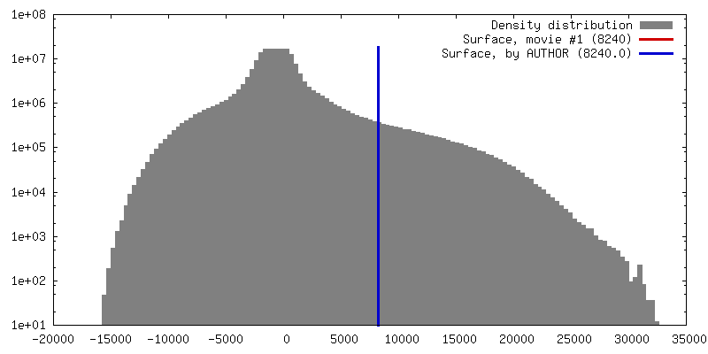
| 密度 |
| ||||||||||||||||||||||||||||||||||||||||||||||||||||||||||||||||||||
| 対称性 | 空間群: 1 | ||||||||||||||||||||||||||||||||||||||||||||||||||||||||||||||||||||
| 詳細 | EMDB XML:
CCP4マップ ヘッダ情報:
| ||||||||||||||||||||||||||||||||||||||||||||||||||||||||||||||||||||
 Z (Sec.)
 Y (Row.)
 X (Col.)


-添付データ
- 試料の構成要素
-全体 : CUS-3 virion, icosahedrally averaged
| 全体 | 名称: CUS-3 virion, icosahedrally averaged |
|---|---|
| 要素 |
|
-超分子 #1000: CUS-3 virion, icosahedrally averaged
| 超分子 | 名称: CUS-3 virion, icosahedrally averaged / タイプ: sample / ID: 1000 / 集合状態: icosahedral / Number unique components: 1 |
|---|
-超分子 #1: Enterobacteria phage CUS-3
| 超分子 | 名称: Enterobacteria phage CUS-3 / タイプ: virus / ID: 1 / NCBI-ID: 539221 / 生物種: Enterobacteria phage CUS-3 / データベース: NCBI / ウイルスタイプ: VIRION / ウイルス・単離状態: SPECIES / ウイルス・エンベロープ: No / ウイルス・中空状態: No |
|---|---|
| 宿主 | 生物種:  |
| ウイルス殻 | Shell ID: 1 / 名称: capsid / 直径: 690 Å / T番号(三角分割数): 7 |
-実験情報
-構造解析
| 手法 | クライオ電子顕微鏡法 |
|---|---|
 解析 解析 | 単粒子再構成法 |
| 試料の集合状態 | particle |
-試料調製
| 濃度 | 10 mg/mL |
|---|---|
| 緩衝液 | pH: 7.6 / 詳細: 10 mM Tris, 10 mM MgCl2 |
| グリッド | 詳細: 400 mesh R2/2 Quantifoil, glow discharged |
| 凍結 | 凍結剤: ETHANE / チャンバー内湿度: 100 % / チャンバー内温度: 90 K / 装置: HOMEMADE PLUNGER / 手法: Blot for 5 sec before plunging. |
- 電子顕微鏡法
| 顕微鏡 | FEI POLARA 300 |
|---|---|
| 温度 | 最低: 89 K / 最高: 91 K / 平均: 90 K |
| アライメント法 | Legacy - 非点収差: Objective lens astigmatism was corrected at the magnification used to collect data. |
| 日付 | 2013年9月1日 |
| 撮影 | カテゴリ: CCD フィルム・検出器のモデル: DIRECT ELECTRON DE-12 (4k x 3k) デジタル化 - サンプリング間隔: 6 µm / 実像数: 419 / 平均電子線量: 23 e/Å2 詳細: The data were collected on the DE12 camera under control of the automated acquisition software, LEGINON. |
| 電子線 | 加速電圧: 200 kV / 電子線源:  FIELD EMISSION GUN FIELD EMISSION GUN |
| 電子光学系 | 照射モード: FLOOD BEAM / 撮影モード: BRIGHT FIELD / Cs: 2.3 mm / 最大 デフォーカス(公称値): 4.08 µm / 最小 デフォーカス(公称値): 0.6 µm / 倍率(公称値): 31000 |
| 試料ステージ | 試料ホルダーモデル: OTHER |
| 実験機器 |  モデル: Tecnai Polara / 画像提供: FEI Company |
-画像解析
| 詳細 | Auto3dem was used for refinement. |
|---|---|
| CTF補正 | 詳細: each micrograph |
| 最終 再構成 | アルゴリズム: OTHER / 解像度のタイプ: BY AUTHOR / 解像度: 6.8 Å / 解像度の算出法: OTHER / ソフトウェア - 名称: Auto3dem, RobEM, autopp 詳細: 28 frames total were collected for each image (35 e-/A2 total dose). Only 15 were used in the final reconstruction (18e-/A2 dose). 使用した粒子像数: 7766 |
