 Movie
Movie Controller

[English] 日本語
 Yorodumi
- EMDB-5938: Electron cryo-microscopy of the Moloney murine leukemia virus fur... -
+ Open data

- Basic information
| Entry | Database: EMDB / ID: EMD-5938 | |||||||||
|---|---|---|---|---|---|---|---|---|---|---|
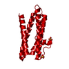
| Title | Electron cryo-microscopy of the Moloney murine leukemia virus furin precursor Env in its native form in complex with 83A25 Fab | |||||||||
 Map data Map data | Reconstruction of the native furin cleavage deficient mutant (R466G/K468G) Env of Moloney murine leukemia virus in complex with the 83A25 Fab | |||||||||
 Sample Sample |
| |||||||||
 Keywords Keywords | Furin precursor / Moloney murine leukemia virus / Env maturation / 83A25 Fab | |||||||||
| Biological species |  Moloney murine leukemia virus / Moloney murine leukemia virus /  | |||||||||
| Method | single particle reconstruction / negative staining / Resolution: 26.0 Å | |||||||||
 Authors Authors | Sjoberg M / Wu SR / Loving R / Rantalainen K / Lindqvist B / Garoff H | |||||||||
 Citation Citation | Journal: Proc Natl Acad Sci U S A / Year: 2014 Title: Furin cleavage of the Moloney murine leukemia virus Env precursor reorganizes the spike structure. Authors: Mathilda Sjöberg / Shang-Rung Wu / Robin Löving / Kimmo Rantalainen / Birgitta Lindqvist / Henrik Garoff /  Abstract: The trimeric Moloney murine leukemia virus Env protein matures by two proteolytic cleavages. First, furin cleaves the Env precursor into the surface (SU) and transmembrane (TM) subunits in the cell ...The trimeric Moloney murine leukemia virus Env protein matures by two proteolytic cleavages. First, furin cleaves the Env precursor into the surface (SU) and transmembrane (TM) subunits in the cell and then the viral protease cleaves the R-peptide from TM in new virus. Here we analyzed the structure of the furin precursor, by cryoelectron microscopy. We transfected 293T cells with a furin cleavage site provirus mutant, R466G/K468G, and produced the virus in the presence of amprenavir to also inhibit the R-peptide cleavage. Although Env incorporation into particles was inhibited, enough precursor could be isolated and analyzed by cryoelectron microscopy to yield a 3D structure at 22 Å resolution. This showed an open cage-like structure like that of the R-peptide precursor and the mature Env described before. However, the middle protrusion of the protomeric unit, so prominently pointing out from the side of the more mature forms of the Env, was absent. Instead, there was extra density in the top protrusion. This suggested that the C-terminal SU domain was associated alongside the receptor binding N-terminal SU domain in the furin precursor. This was supported by mapping with a SU C-terminal domain-specific antigen binding fragment. We concluded that furin cleavage not only separates the subunits and liberates the fusion peptide at the end of TM but also allows the C-terminal domain to relocate into a peripheral position. This conformational change might explain how the C-terminal domain of SU gains the potential to undergo disulfide isomerization, an event that facilitates membrane fusion. | |||||||||
| History |
|
- Structure visualization
| Movie |
 Movie viewer Movie viewer |
|---|---|
| Structure viewer | EM map: SurfViewMolmilJmol/JSmol |
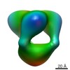
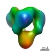
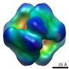
| Supplemental images |

- Downloads & links
-EMDB archive
| Map data | emd_5938.map.gz | 800.1 KB | EMDB map data format | |
|---|---|---|---|---|
| Header (meta data) | emd-5938-v30.xmlemd-5938.xml | 12.1 KB 12.1 KB | Display Display | EMDB header |
| Images | 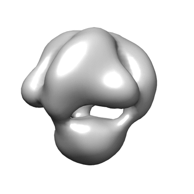 400_5938.gif 400_5938.gif 80_5938.gif 80_5938.gif | 24.6 KB 2.8 KB | ||
| Archive directory |  http://ftp.pdbj.org/pub/emdb/structures/EMD-5938ftp://ftp.pdbj.org/pub/emdb/structures/EMD-5938 http://ftp.pdbj.org/pub/emdb/structures/EMD-5938ftp://ftp.pdbj.org/pub/emdb/structures/EMD-5938 | HTTPS FTP |
-Related structure data

-Links
| EMDB pages | EMDB (EBI/PDBe) / EMDataResource |
|---|
-Map
| File | Download / File: emd_5938.map.gz / Format: CCP4 / Size: 1001 KB / Type: IMAGE STORED AS FLOATING POINT NUMBER (4 BYTES) | ||||||||||||||||||||||||||||||||||||||||||||||||||||||||||||||||||||
|---|---|---|---|---|---|---|---|---|---|---|---|---|---|---|---|---|---|---|---|---|---|---|---|---|---|---|---|---|---|---|---|---|---|---|---|---|---|---|---|---|---|---|---|---|---|---|---|---|---|---|---|---|---|---|---|---|---|---|---|---|---|---|---|---|---|---|---|---|---|
| Annotation | Reconstruction of the native furin cleavage deficient mutant (R466G/K468G) Env of Moloney murine leukemia virus in complex with the 83A25 Fab | ||||||||||||||||||||||||||||||||||||||||||||||||||||||||||||||||||||
| Projections & slices | Image control
Images are generated by Spider. | ||||||||||||||||||||||||||||||||||||||||||||||||||||||||||||||||||||
| Voxel size | X=Y=Z: 3.5 Å | ||||||||||||||||||||||||||||||||||||||||||||||||||||||||||||||||||||
| Density |
| ||||||||||||||||||||||||||||||||||||||||||||||||||||||||||||||||||||
| Symmetry | Space group: 1 | ||||||||||||||||||||||||||||||||||||||||||||||||||||||||||||||||||||
| Details | EMDB XML:
CCP4 map header:
| ||||||||||||||||||||||||||||||||||||||||||||||||||||||||||||||||||||
 Z (Sec.)
 Y (Row.)
 X (Col.)


-Supplemental data
- Sample components
-Entire : Native form of furin cleavage deficient mutant (R466G/K468G) Env ...
| Entire | Name: Native form of furin cleavage deficient mutant (R466G/K468G) Env of Moloney murine leukemia virus in complex with 83A25 Fab |
|---|---|
| Components |
|
-Supramolecule #1000: Native form of furin cleavage deficient mutant (R466G/K468G) Env ...
| Supramolecule | Name: Native form of furin cleavage deficient mutant (R466G/K468G) Env of Moloney murine leukemia virus in complex with 83A25 Fab type: sample / ID: 1000 Details: Affinity purified, gradient separated protein in 0.05% Triton X-100 Oligomeric state: trimer / Number unique components: 2 |
|---|---|
| Molecular weight | Experimental: 600 KDa / Theoretical: 400 KDa / Method: Estimated from Blue native PAGE |
-Macromolecule #1: gp90
| Macromolecule | Name: gp90 / type: protein_or_peptide / ID: 1 / Name.synonym: Furin precursor / Number of copies: 3 / Oligomeric state: Trimer / Recombinant expression: No / Database: NCBI |
|---|---|
| Source (natural) | Organism: Moloney murine leukemia virus |
| Molecular weight | Experimental: 500 KDa / Theoretical: 270 KDa |
-Macromolecule #2: monoclonal antibody 83A25 Fab
| Macromolecule | Name: monoclonal antibody 83A25 Fab / type: protein_or_peptide / ID: 2 / Details: 2-3 Fab molecules bind to the gp90 trimer. / Number of copies: 3 / Oligomeric state: monomer / Recombinant expression: No / Database: NCBI |
|---|---|
| Source (natural) | Organism: |
| Molecular weight | Experimental: 50 KDa / Theoretical: 50 KDa |
-Experimental details
-Structure determination
| Method | negative staining |
|---|---|
 Processing Processing | single particle reconstruction |
| Aggregation state | particle |
-Sample preparation
| Concentration | 0.1 mg/mL |
|---|---|
| Buffer | pH: 7.4 / Details: 50 mM HEPES, 100 mM NaCl, 1.8 mM CaCl2, pH 7.4 |
| Staining | Type: NEGATIVE Details: The specimen was stained with 2% uranyl acetate (UA). |
| Grid | Details: 400 mesh regular continuous carbon grid, glow discharged |
| Vitrification | Cryogen name: NONE / Instrument: OTHER |
- Electron microscopy
| Microscope | JEOL 2100F |
|---|---|
| Alignment procedure | Legacy - Astigmatism: Objective lens astigmatism was corrected using online FFT. |
| Date | Jul 5, 2013 |
| Image recording | Category: CCD / Film or detector model: TVIPS TEMCAM-F416 (4k x 4k) / Digitization - Sampling interval: 3.5 µm / Number real images: 60 / Average electron dose: 9 e/Å2 / Bits/pixel: 14 |
| Electron beam | Acceleration voltage: 200 kV / Electron source:  FIELD EMISSION GUN FIELD EMISSION GUN |
| Electron optics | Calibrated magnification: 43200 / Illumination mode: SPOT SCAN / Imaging mode: BRIGHT FIELD / Cs: 2.0 mm / Nominal defocus max: 2.072 µm / Nominal defocus min: 2.048 µm / Nominal magnification: 43200 |
| Sample stage | Specimen holder model: OTHER |
+Image processing
-Atomic model buiding 1
| Initial model | PDB ID: |
|---|---|
| Software | Name: Chimera |
| Details | Atomic structure of the Fv fragment of the monoclonal Ab 7E2 against the Paracoccus denitrificans cytochrome c oxidase was fitted into the density map. |
| Refinement | Space: REAL / Protocol: RIGID BODY FIT |

